

Rapid release of plasmid DNA from surfaces coated with polyelectrolyte multilayers promoted by the application of electrochemical potentials
- PMID: 22551230
- PMCID: PMC3359390
- DOI: 10.1021/am3003632
Rapid release of plasmid DNA from surfaces coated with polyelectrolyte multilayers promoted by the application of electrochemical potentials
Abstract
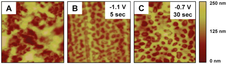
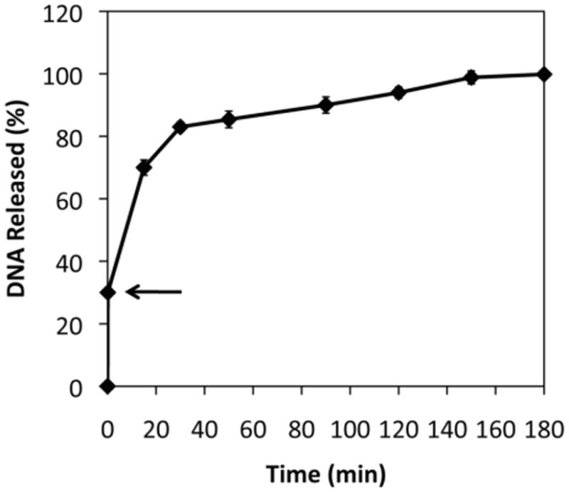
We report an approach to the rapid release of DNA based on the application of electrochemical potentials to surfaces coated with polyelectrolyte-based thin films. We fabricated multilayered polyelectrolyte films (or "polyelectrolyte multilayers", PEMs) using plasmid DNA and a model hydrolytically degradable cationic poly(β-amino ester) (polymer 1) on stainless steel substrates using a layer-by-layer approach. The application of continuous reduction potentials in the range of -1.1 to -0.7 V (vs a Ag/AgCl electrode) to film-coated electrodes in PBS at 37 °C resulted in the complete release of DNA over a period of 1-2 min. Film-coated electrodes incubated under identical conditions in the absence of applied potentials required 1-2 days for complete release. Control over the magnitude of the applied potential provided control over the rate at which DNA was released. The results of these and additional physical characterization experiments are consistent with a mechanism of film disruption that is promoted by local increases in pH at the film/electrode interface (resulting from electrochemical reduction of water or dissolved oxygen) that disrupt ionic interactions in these materials. The results of cell-based experiments demonstrated that DNA was released in a form that remains intact and able to promote transgene expression in mammalian cells. Finally, we demonstrate that short-term (i.e., non-continuous) electrochemical treatments can also be used to promote faster film erosion (e.g., over 1-2 h) once the potential is removed. Past studies demonstrate that PEMs fabricated using polymer 1 can promote surface-mediated transfection of cells and tissues in vitro and in vivo. With further development, the electrochemical approaches reported here could thus provide new methods for the rapid, triggered, or spatially patterned transfer of DNA (or other agents) from surfaces of interest in a variety of fundamental and applied contexts.
Figures


Similar articles
-
Degradable polyelectrolyte multilayers that promote the release of siRNA.Langmuir. 2011 Jun 21;27(12):7868-76. doi: 10.1021/la200815t. Epub 2011 May 16. Langmuir. 2011. PMID: 21574582 Free PMC article.
-
Release of DNA from polyelectrolyte multilayers fabricated using 'charge-shifting' cationic polymers: tunable temporal control and sequential, multi-agent release.J Control Release. 2010 Nov 20;148(1):91-100. doi: 10.1016/j.jconrel.2010.07.112. Epub 2010 Jul 30. J Control Release. 2010. PMID: 20678530 Free PMC article.
-
Characterization of degradable polyelectrolyte multilayers fabricated using DNA and a fluorescently-labeled poly(β-amino ester): shedding light on the role of the cationic polymer in promoting surface-mediated gene delivery.Biomacromolecules. 2012 Feb 13;13(2):542-52. doi: 10.1021/bm2016338. Epub 2012 Jan 6. Biomacromolecules. 2012. PMID: 22224541 Free PMC article.
-
Multilayered films fabricated from plasmid DNA and a side-chain functionalized poly(beta-amino ester): surface-type erosion and sequential release of multiple plasmid constructs from surfaces.Langmuir. 2007 Oct 23;23(22):11139-46. doi: 10.1021/la702021s. Epub 2007 Sep 22. Langmuir. 2007. PMID: 17887783
-
Multilayered polyelectrolyte assemblies as platforms for the delivery of DNA and other nucleic acid-based therapeutics.Adv Drug Deliv Rev. 2008 Jun 10;60(9):979-99. doi: 10.1016/j.addr.2008.02.010. Epub 2008 Mar 4. Adv Drug Deliv Rev. 2008. PMID: 18395291 Free PMC article. Review.
Cited by
-
Programmable multi-DNA release from multilayered polyelectrolytes using gigahertz nano-electromechanical resonator.J Nanobiotechnology. 2019 Aug 6;17(1):86. doi: 10.1186/s12951-019-0518-7. J Nanobiotechnology. 2019. PMID: 31387581 Free PMC article.
-
Biomaterial substrate modifications that influence cell-material interactions to prime cellular responses to nonviral gene delivery.Exp Biol Med (Maywood). 2019 Feb;244(2):100-113. doi: 10.1177/1535370218821060. Epub 2019 Jan 8. Exp Biol Med (Maywood). 2019. PMID: 30621454 Free PMC article. Review.
-
Electrical Field-Assisted Gene Delivery from Polyelectrolyte Multilayers.Polymers (Basel). 2020 Jan 6;12(1):133. doi: 10.3390/polym12010133. Polymers (Basel). 2020. PMID: 31935814 Free PMC article.
-
Phenylboronic Acid-Functionalized Layer-by-Layer Assemblies for Biomedical Applications.Polymers (Basel). 2017 May 31;9(6):202. doi: 10.3390/polym9060202. Polymers (Basel). 2017. PMID: 30970879 Free PMC article. Review.
-
DNA Computing Systems Activated by Electrochemically-triggered DNA Release from a Polymer-brush-modified Electrode Array.Electroanalysis. 2017 Feb;29(2):398-408. doi: 10.1002/elan.201600389. Epub 2016 Aug 5. Electroanalysis. 2017. PMID: 29379265 Free PMC article.
References
Publication types
MeSH terms
Substances
Grants and funding
LinkOut - more resources
Full Text Sources
Miscellaneous
