. 2012 May 2;14(3):R106.
doi: 10.1186/ar3831.

Efficacy of an immunotoxin to folate receptor beta in the intra-articular treatment of antigen-induced arthritis

Affiliations

Efficacy of an immunotoxin to folate receptor beta in the intra-articular treatment of antigen-induced arthritis

Taku Nagai et al. Arthritis Res Ther. .

Abstract

Introduction: We previously demonstrated that synovial sublining macrophages express folate receptor beta (FRβ). The aim of this study was to evaluate the efficacy of intra-articular administration of a recombinant immunotoxin to FRβ for treating rat antigen-induced arthritis.

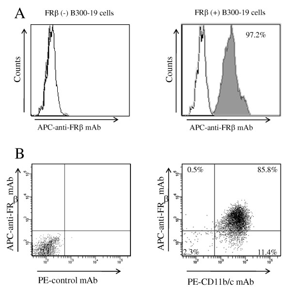
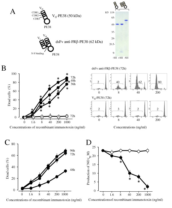
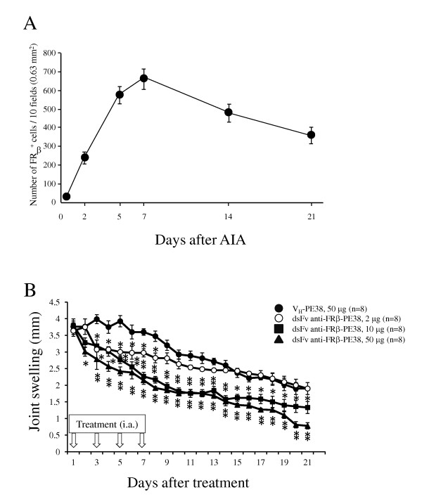
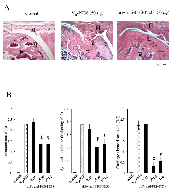
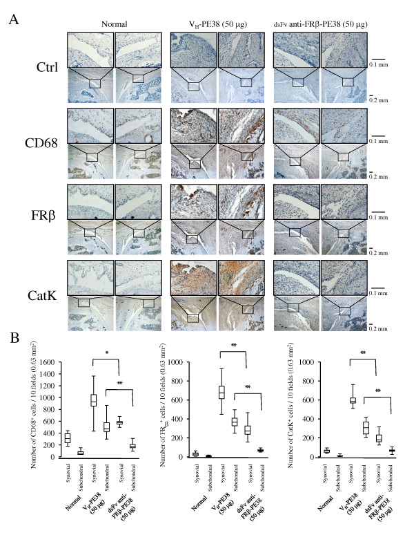
Methods: A monoclonal antibody (mAb) to rat FRβ was produced by immunizing mice with B300-19 cells (murine pre-B cells) transfected with the rat FRβ gene. Recombinant immunotoxin was prepared by conjugating the Fv portion of the anti-rat FRβ mAb heavy chain with a truncated Pseudomonas exotoxin A and the Fv portion of the anti-rat FRβ mAb light chain. Antigen-induced arthritis was induced through intra-articular injection of methylated bovine serum albumin (mBSA) after two subcutaneous injections of mBSA and complete Freund's adjuvant. Immunotoxin was intra-articularly injected into the arthritis joint every other day for seven days after arthritis onset. Joint swelling was measured and histological scores of inflammation, synovial thickness, cartilage, and bone destruction were determined. Immunohistochemistry was performed to detect osteoclast and osteoclast precursor FRβ-expressing macrophages and cathepsin K-positive cells on day 21.

Results: Intra-articular administration of the immunotoxin attenuated joint swelling (61% suppression; P < 0.01 compared to the control on day 21) and improved histological findings, particularly cartilage and bone destruction (scores of rats treated with control versus the immunotoxin: 2.2 versus 0.5; P < 0.01), by reducing the number of FRβ-expressing macrophages and cathepsin K-positive cells.

Conclusions: Intra-articular administration of an immunotoxin to FRβ is effective for improving rat antigen-induced arthritis.


Figures

Figure 1
Figure 1
Reactivity of an anti-rat FRβ mAb with FRβ-expressing cells. (A) Non-transfected B300-19 cells (left) and rat FRβ gene-transfected B300-19 cells. (right) were stained using an anti-rat FRβ (4A67) mAb (black pattern) or isotype-matched irrelevant mAb (white pattern). Stained cells were analyzed using flow cytometry. Data are representative of three separate experiments. (B) TGC-elicited macrophages were double-stained using an anti-FRβ mAb (4A67) and PE-conjugated CD11b/c mAbs (OX42) and analyzed using flow cytometry. Numbers represent the percentage of cells within designated gates. Data are representative of three separate experiments. FRβ, folate receptor β; mAb, monoclonal antibody; TGC, thioglycollate.
Figure 2
Figure 2
Reactivity of an anti-rat FRβ mAb with rat lung and liver tissues. Lung and liver tissues from normal rats were stained using antibodies against CD68, FRβ, or E-cadherin. Photographs are representative of CD68-, FRβ-, and E-cadherin staining in lung or liver tissues of three rats per group. Note that FRβ-positive cells were observed in the liver but not in the lung. CD68- positive cells and E-cadherin-positive cells (indicating the presence of epithelial cells) were observed in both tissues; original magnification was ×400. FRβ, folate receptor β; mAb, monoclonal antibody.
Figure 3
Figure 3
Effects of anti-FRβ immunotoxin on cell death induction and NO production of FRβ-expressing cells. (A)The schema shows the construction of VH-PE38 and dsFv anti-FRβ-PE38. The purity of VH-PE38 and dsFv anti-FRβ-PE38 were analyzed using sodium dodecyl sulphate- 6% to 15% gradient gel electrophoresis in non-reducing (-SH) and reducing conditions (+SH). Molecular weight markers are shown at left. (B) Rat FRβ-transfected B300-19 cells were cultured at the indicated concentrations of VH-PE38 (open triangle) for 72 hours or at the indicated concentrations of dsFv anti-FRβ-PE38 (filled circle) for 36, 48, or 72 hours. Non-transfected B300-19 cells were also cultured at the indicated concentrations of dsFv anti-FRβ-PE38 (open circle) for 36, 48, or 72 hours. The number of dead cells was measured using propidium iodide staining and flow cytometric analysis, and the percentage of dead cells was calculated as the number of dead cells divided by total cell number. Values are the mean ± SEM of induced cell death from three separate experiments. The right panels are representative of propidium iodide staining patterns at the indicated concentrations of VH-PE38 or dsFv anti-FRβ-PE38. Values in panels show the percentages of dead cells. (C) Thioglycollate-elicited macrophages were cultured at the indicated concentrations of VH-PE38 or dsFv anti-FRβ-PE38 for 48, 72, or 96 hours. Induced cell death (%) was determined by subtracting the percentage of dead cells induced by VH-PE38 from that induced by dsFv anti-FRβ-PE38 in each sample. Values are the mean ± SEM of induced cell death of three separate experiments. (D) Thioglycollate-elicited macrophages were cultured at the indicated concentrations of VH-PE38 (open circle) or dsFv anti-FRβ-PE38 (filled circle) for 24 hours and stimulated using IFN-γ and LPS for an additional 48 hours. NO in the supernatants was measured using the Griess reaction. Values are the mean ± SEM of three separate experiments. * P < 0.05 compared to the VH-PE38 group. FRβ, folate receptor β; IFN-γ, interferon-γ; LPS, Lipopolysaccharide; SEM, standard error of the mean.
Figure 4
Figure 4
Effects of anti-FRβ immunotoxin on knee joint swelling in rat AIA. (A) FRβ-expressing macrophages in synovium of the arthritis rat were examined on the indicated days after AIA onset as described in Methods. Values are the mean ± SEM of knee joints from four rats per group. (B) AIA was induced using intra-articular injection of methylated BSA as described in Methods. Arthritic rats were injected in the left knee joint with 50 μg of VH-PE38 (filled circle) or with 2 µg (open circle), 10 µg (filled square) or 50 μg (filled triangle) of dsFv anti-FRβ-PE38 on days one, three, five and seven. Knee joint swelling was measured for each rat by measuring the difference in diameter between the arthritic left knee and the normal right knee. Values are the mean ± SEM of knee joint swelling from eight rats per group. *P <0.05 and **P <0.01 compared to the VH-PE38-treated group. AIA, antigen-induced arthritis; BSA, bovine serum albumin; FRβ, folate receptor β; SEM, standard error of the mean.
Figure 5
Figure 5
Effects of an anti-FRβ immunotoxin on the histology of knee joints in rat AIA. (A)Photographs are representative of knee joints from eight rats per group. Knee joints from normal rats and AIA rats treated with 50 µg of VH-PE38 or dsFv anti-FRβ-PE38 were stained using H & E. Note that a knee joint from an AIA rat treated using dsFv anti-FRβ-PE38 showed less cartilage/bone destruction compared to a rat treated using VH-PE38. Letters S, C, B, and E represent synovium, cartilage, bone, and erosion. Original magnification was ×100. (B)Histological scores were recorded for inflammation, synovial membrane thickening, and cartilage/bone destruction. Data are presented as the mean ± SEM of each histological score from eight rats per group. *P < 0.05 and **P < 0.01 compared to the group treated using VH-PE38. AIA, antigen-induced arthritis; FRβ, folate receptor β; H & E, haematoxylin and eosin; SEM, standard error of the mean.
Figure 6
Figure 6
CD68-, FRβ-, and cathepsin K-positive cells in the knee joints of AIA rats treated using an anti-FRβ immunotoxin. (A) Knee joints from normal rats and AIA rats treated with 50 µg of VH-PE38 or dsFv anti-FRβ-PE38 were stained with isotype-matched controls (a mixture of mouse IgG1 and IgM mAbs, and rabbit IgG), antibodies against CD68, FRβ, or cathepsin K, and positive cells were counted. Photographs are representative of negative control and CD68-, FRβ-, and cathepsin K-positive cells in the knee joints of eight rats per group. Note that the synovium from AIA rats treated using dsFv anti-FRβ-PE38 showed fewer CD68-, FRβ-, and cathepsin K-positive cells compared to those treated using VH-PE38. Original magnifications were ×100 and ×400. (B)Numbers of CD68-, FRβ-, and cathepsin K-positive cells were counted in the knee joints of each group. Data from eight rats per group are presented as box plots, where the boxes represent the 25th to 75th percentiles, the lines within the boxes represent the median, and the lines outside the boxes represent the 10th and 90th percentiles. *P < 0.05 and **P < 0.01 compared to the group treated using VH-PE38. AIA, antigen-induced arthritis; FRβ, folate receptor β; Ig, immunoglobulin.

Similar articles

Cited by

References

    1. Szekanecz Z, Koch AE. Macrophages and their products in rheumatoid arthritis. Curr Opin Rheumatol. 2007;19:289–295. doi: 10.1097/BOR.0b013e32805e87ae. - DOI - PubMed
    1. McHugh KP, Shen Z, Crotti TN, Flannery MR, O'Sullivan RP, Purdue PE, Goldring SR. The role of cell-substrate interaction in regulating osteoclast activation: potential implications in targeting bone loss in rheumatoid arthritis. Ann Rheum Dis. 2010;69(Suppl 1):i83–85. doi: 10.1136/ard.2009.120188. - DOI - PubMed
    1. Mulherin D, Fitzgerald O, Bresnihan B. Synovial tissue macrophage populations and articular damage in rheumatoid arthritis. Arthritis Rheum. 1996;39:115–124. doi: 10.1002/art.1780390116. - DOI - PubMed
    1. Tak PP, Smeets TJ, Daha MR, Kluin PM, Meijers KA, Brand R, Meinders AE, Breedveld C. Analysis of the synovial cell infiltrate in early rheumatoid synovial tissue in relation to local disease activity. Arthritis Rheum. 1997;40:217–225. doi: 10.1002/art.1780400206. - DOI - PubMed
    1. Bresnihan B, Pontifex E, Thurlings RM, Vinkenoog M, El-Gabalawy H, Fearon U, Fitzgerald O, Gerlag DM, Rooney T, van de Sande MG, Veale D, Vos K, Tak PP. Synovial tissue sublining CD68 expression is a biomarker of therapeutic response in rheumatoid arthritis clinical trials: consistency across centers. J Rheumatol. 2009;36:1800–1802. doi: 10.3899/jrheum.090348. - DOI - PubMed

Publication types

MeSH terms