. 2012 Jul;33(21):5287-96.
doi: 10.1016/j.biomaterials.2012.04.010. Epub 2012 Apr 30.

Characterization of natural, decellularized and reseeded porcine tooth bud matrices

Affiliations

Characterization of natural, decellularized and reseeded porcine tooth bud matrices

Samantha B Traphagen et al. Biomaterials. 2012 Jul.

Abstract

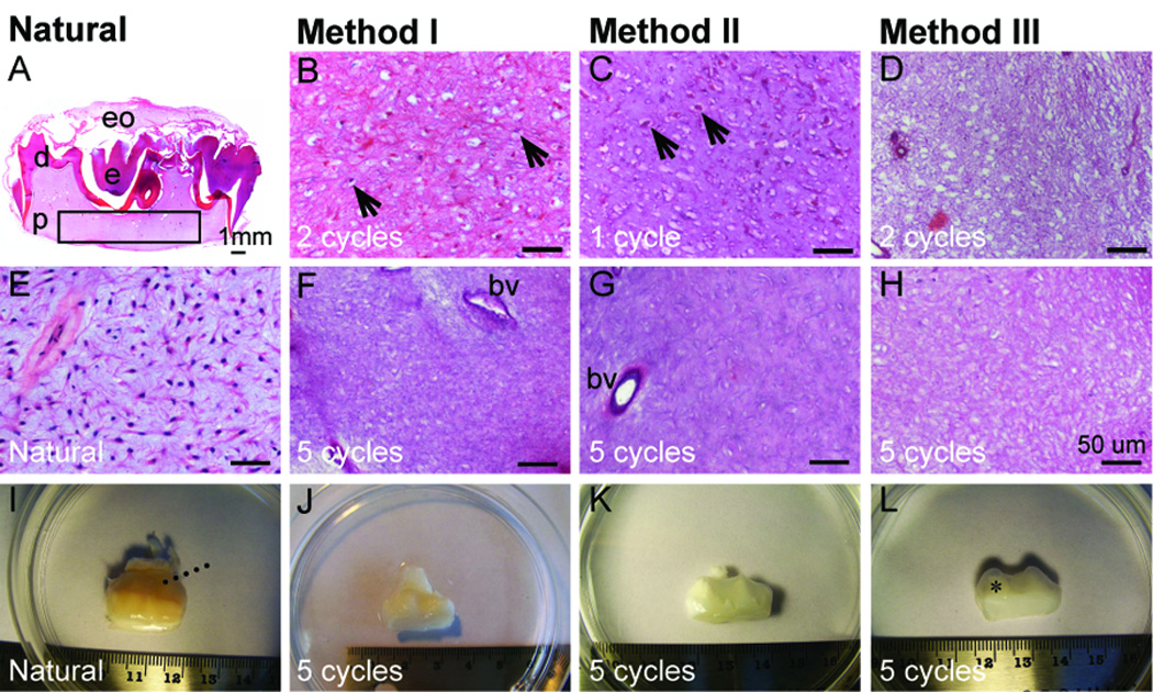
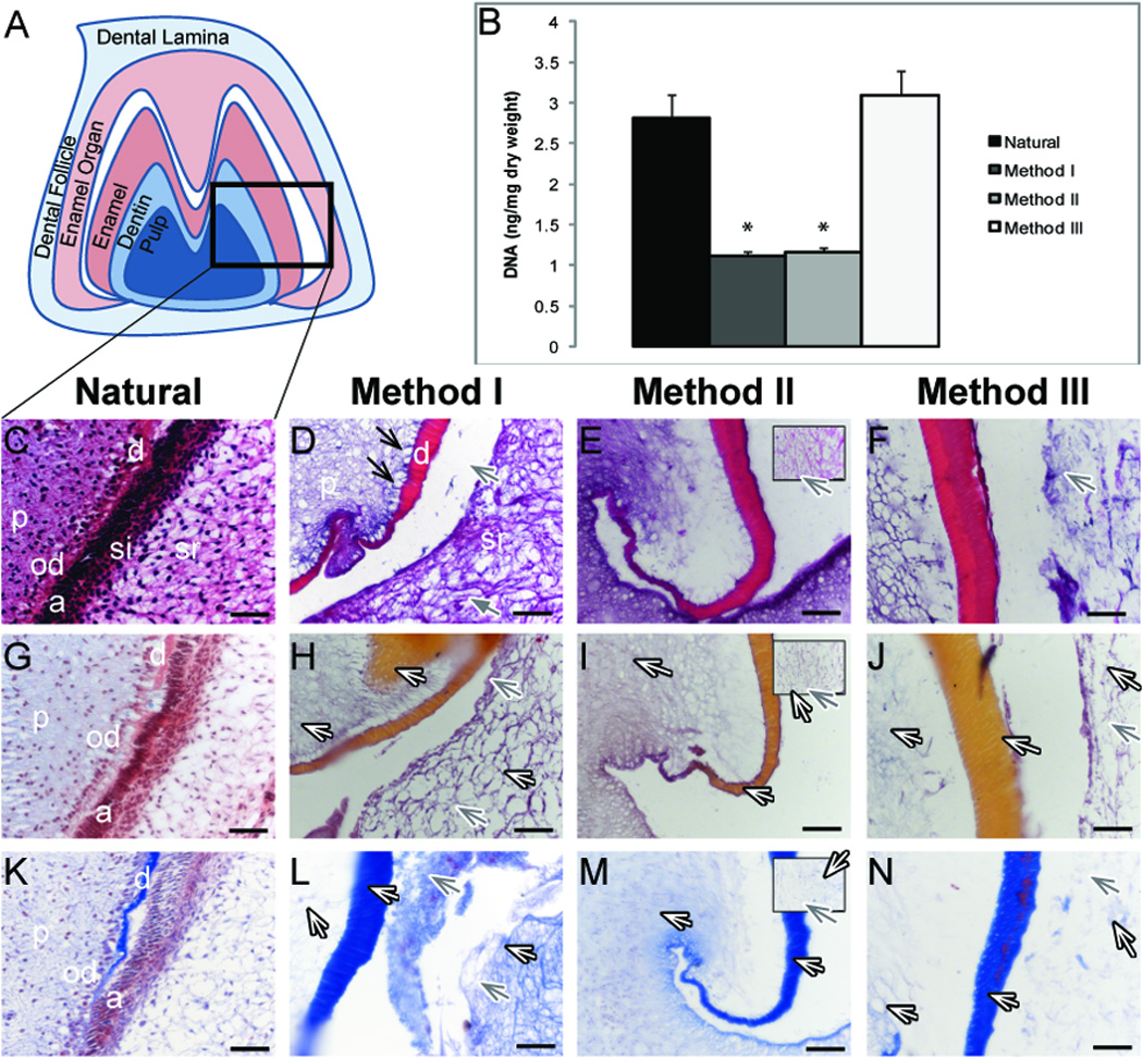
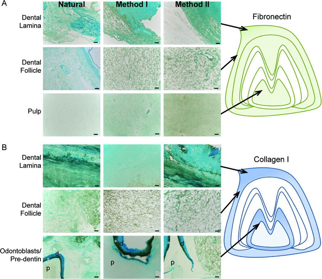
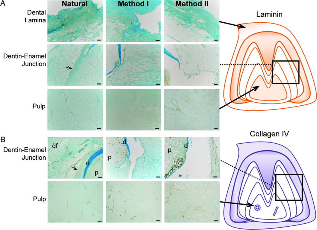
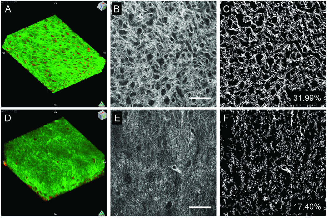
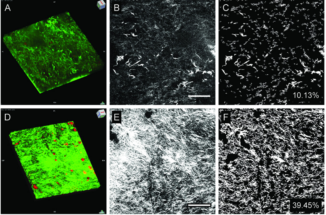
Dental tissue engineering efforts have yet to identify scaffolds that instruct the formation of bioengineered teeth of predetermined size and shape. Here we investigated whether extracellular matrix (ECM) molecules present in natural tooth scaffolds can provide insight on how to achieve this goal. We describe methods to effectively decellularize and demineralize porcine molar tooth buds, while preserving natural ECM protein gradients. Natural tooth ECM composition was assessed using histological and immunohistochemical (IHC) analyses of fibrillar and basement membrane proteins. Our results showed that Collagen I, Fibronectin, Collagen IV, and Laminin gradients were detected in natural tooth tissues, and retained in decellularized samples. Second harmonic generation (SHG) image analysis and 3D reconstructions were used to show that natural tooth tissue exhibited higher collagen fiber density, and less oriented and less organized collagen fibers, as compared to decellularized tooth tissue. We also found that reseeded decellularized tooth scaffolds exhibited distinctive collagen content and organization as compared to decelluarized scaffolds. Our results show that SHG allows for quantitative assessment of ECM features that are not easily characterized using traditional histological analyses. In summary, our results demonstrate the potential for natural decellularized molar tooth ECM to instruct dental cell matrix synthesis, and lay the foundation for future use of biomimetic scaffolds for dental tissue engineering applications.


Figures

Figure 1
Figure 1. Comparison of natural and decellularized tooth buds
(A, E) H&E stained natural M2 molar tooth section reveals highly cellularized dental pulp (p) (boxed region). H&E stained sectioned pulp after various Method I (B, F), Method II (C, G), and Method III (D, H) treatments. (I–L) Low mag view of teeth after various treatments. Black arrows indicate nuclei, dashed lines indicate the dental sac, and * denotes tooth cusps. Abbreviations: bv, blood vessel; d, dentin, e, enamel; eo, enamel organ. Scale bars: (A) 1.0 mm; (B–H) 50 microns.
Figure 2
Figure 2. Decellularized tooth samples
(A). Schematic of M3 tooth. B) DNA content before and after decellularization. H&E (C–F), Movat’s Pentachrome (G–J) and Masson’s Trichrome (K–N) stained paraffin sections. Method II boxed inlays display stellate reticulum (E, I, M). Efficiently removed odontoblasts (D, black arrows) and ameloblasts (D, gray arrows), and preserved collagen (white arrows). Abbreviations: a ameloblasts; d, dentin; p, pulp; od, odontoblasts; si, stratum intermedium; sr, stellate reticulum. * = p < 0.0001. Scale bar = 50 microns.
Figure 3
Figure 3. IHC analyses of fibrillar ECM protein expression
(A) Fibronectin and (B) Collagen I expression in Natural, Method I, or Method II treated teeth. Schematic representations are shown to the right. Strong collagen I expression was observed in the pre-dentin region (*) between the pulp (p) and dentin (d). Scale = 50 microns.
Figure 4
Figure 4. IHC analyses of basement membrane ECM protein expression
(A) Laminin and (B) Collagen IV expression in Natural, Method I, or Method II treated teeth. Schematic representations are shown to the right. Vascular and nerve tissues expressed both basement membrane proteins. Abbreviations: d, dentin; p, pulp; dental follicle, df. Scale = 50 microns.
Figure 5
Figure 5. Non-linear microscopy of natural and decellularized M2 molar pulp
TPEF (red pseudocolor) and SHG (green pseudocolor) reconstructions of natural (A) and Method I treated (D) tooth scaffolds (238×238×64 µm3 tissue volumes). Representative natural (B) and decellularized (E) tooth scaffolds. Thresholded natural (C) and decellularized (H) images were used to assess collagen fiber density and organization. Scale = 50 microns.
Figure 6
Figure 6. Non-linear microscopy of re-seeded tooth scaffolds after four weeks of culture
SHG (green pseudocolor) and TPEF (red pseudocolor) reconstructions of cultured unseeded (A), and cultured dental cell seeded (D) decellularized scaffolds (238×238×33 µm3 tissue volumes). Representative images of cultured unseeded (B) and mes-seeded (E) scaffolds. Collagen fiber densities were determined from thresholded images (C, H). Scale bars = 50 microns.

Similar articles

Cited by

References

    1. Honda MJ, Fong H, Iwatsuki S, Sumita Y, Sarikaya M. Tooth-forming potential in embryonic and postnatal tooth bud cells. Med Mol Morphol. 2008;41:183–192. - PubMed
    1. Li YC, Jin F, Du Y, Ma ZW, Li F, Wu G, et al. Cementum and periodontal ligament-like tissue formation induced using bioengineered dentin. Tissue Eng Part A. 2008;14:1731–1742. - PubMed
    1. Nakao K, Morita R, Saji Y, Ishida K, Tomita Y, Ogawa M, et al. The development of a bioengineered organ germ method. Nat Methods. 2007;4:227–230. - PubMed
    1. Scheller EL, Krebsbach PH, Kohn DH. Tissue engineering: state of the art in oral rehabilitation. J Oral Rehabil. 2009;36:368–389. - PMC - PubMed
    1. Du C, Fan DM, Sun Z, Fan YW, Lakshminarayanan R, Moradian-Oldak J. Immunogold labeling of amelogenin in developing porcine enamel revealed by field emission scanning electron microscopy. 9th International Conference on the Chemistry and Biology of Mineralized Tissues; Karger; Austin, TX. 2007. pp. 207–211. - PMC - PubMed

Publication types