. 2013 Mar;20(3):274-82.
doi: 10.1038/gt.2012.38. Epub 2012 May 3.

MRI roadmap-guided transendocardial delivery of exon-skipping recombinant adeno-associated virus restores dystrophin expression in a canine model of Duchenne muscular dystrophy

Affiliations

MRI roadmap-guided transendocardial delivery of exon-skipping recombinant adeno-associated virus restores dystrophin expression in a canine model of Duchenne muscular dystrophy

I M Barbash et al. Gene Ther. 2013 Mar.

Abstract

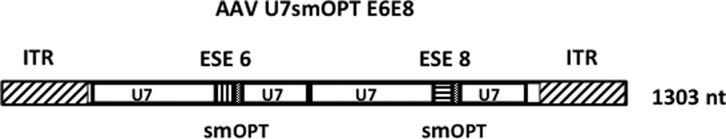
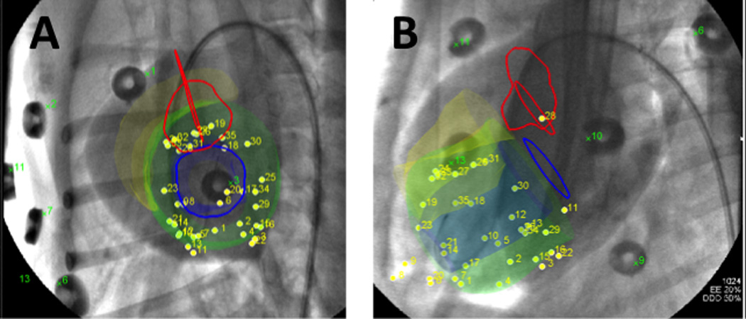
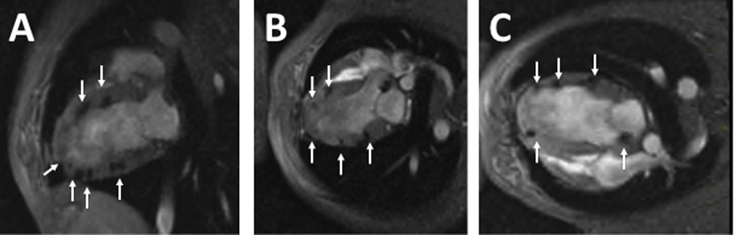
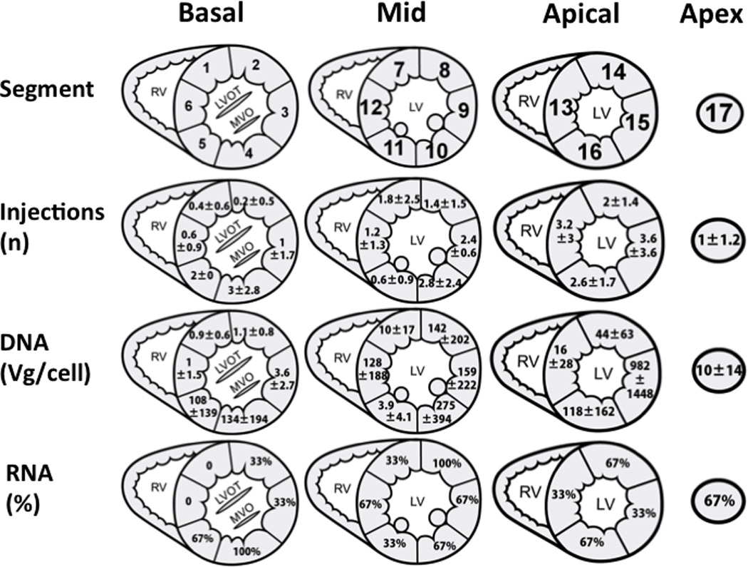
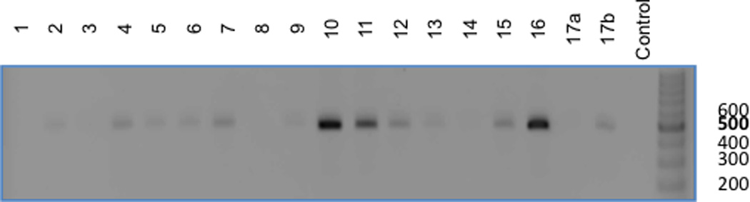
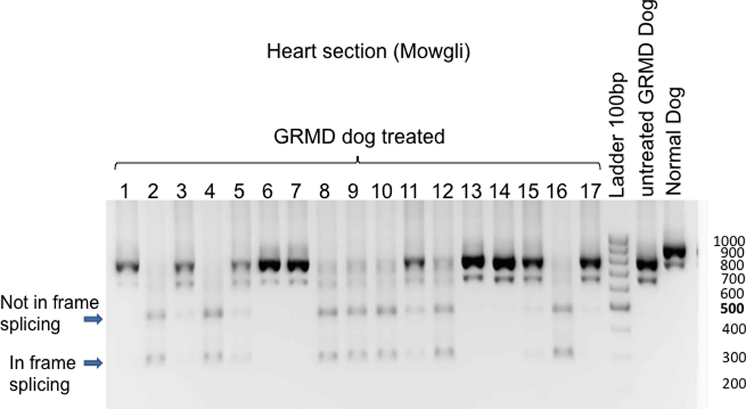
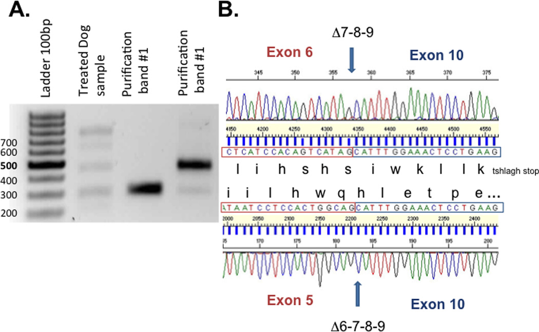
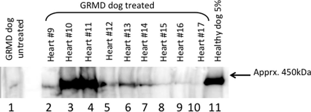
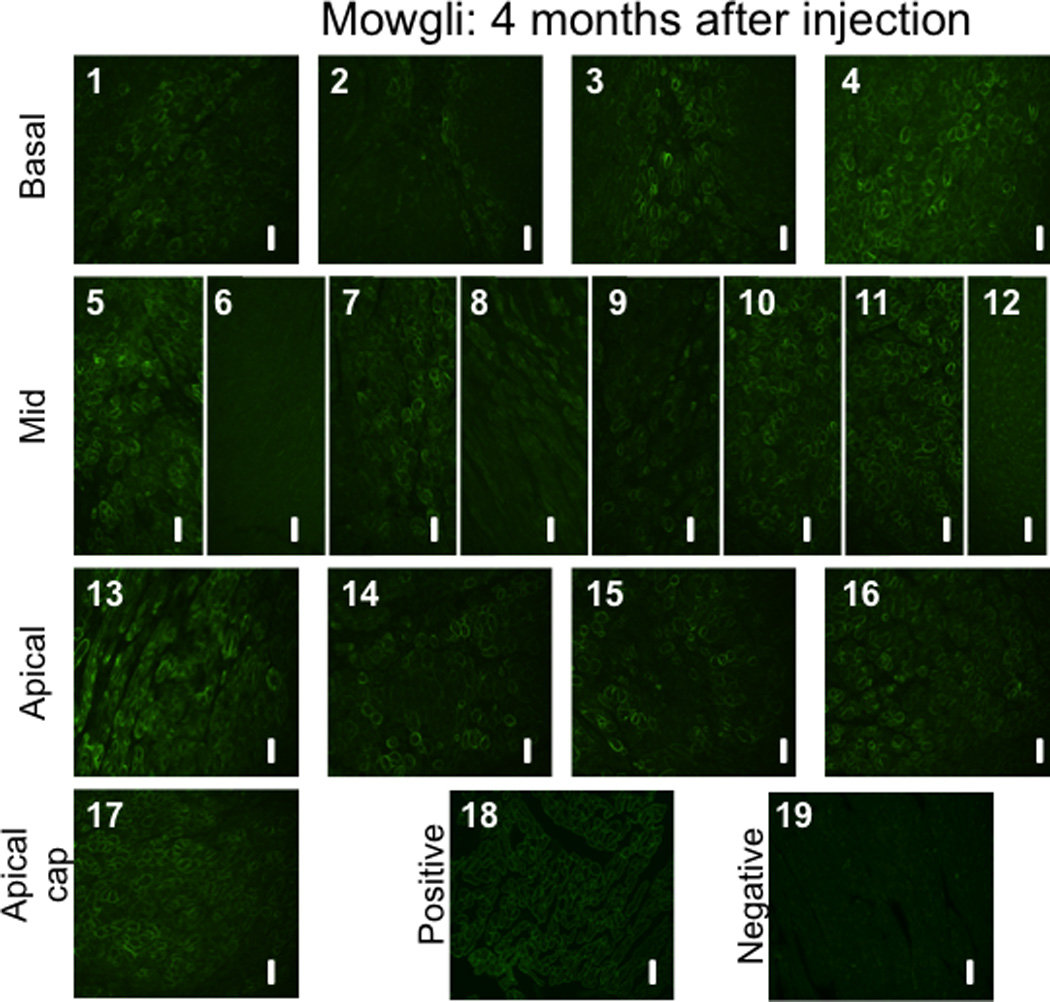
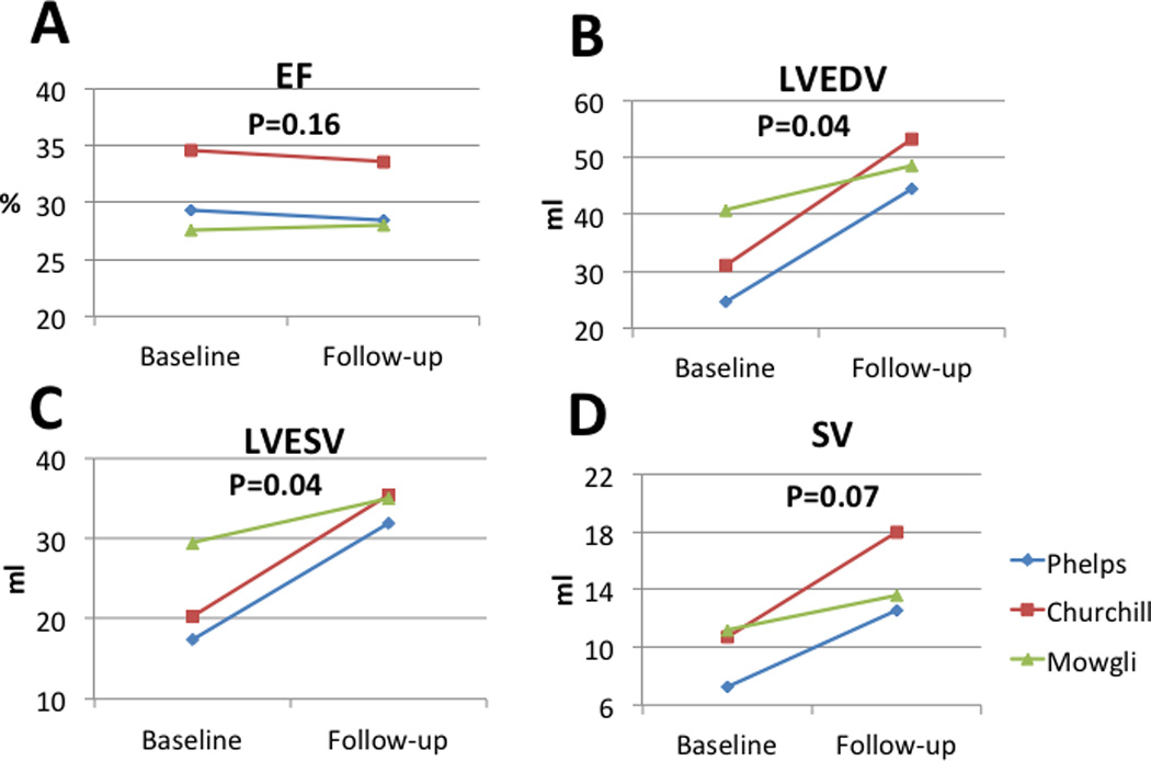
Duchenne muscular dystrophy (DMD) cardiomyopathy patients currently have no therapeutic options. We evaluated catheter-based transendocardial delivery of a recombinant adeno-associated virus (rAAV) expressing a small nuclear U7 RNA (U7smOPT) complementary to specific cis-acting splicing signals. Eliminating specific exons restores the open reading frame resulting in translation of truncated dystrophin protein. To test this approach in a clinically relevant DMD model, golden retriever muscular dystrophy (GRMD) dogs received serotype 6 rAAV-U7smOPT via the intracoronary or transendocardial route. Transendocardial injections were administered with an injection-tipped catheter and fluoroscopic guidance using X-ray fused with magnetic resonance imaging (XFM) roadmaps. Three months after treatment, tissues were analyzed for DNA, RNA, dystrophin protein, and histology. Whereas intracoronary delivery did not result in effective transduction, transendocardial injections, XFM guidance, enabled 30±10 non-overlapping injections per animal. Vector DNA was detectable in all samples tested and ranged from <1 to >3000 vector genome copies per cell. RNA analysis, western blot analysis, and immunohistology demonstrated extensive expression of skipped RNA and dystrophin protein in the treated myocardium. Left ventricular function remained unchanged over a 3-month follow-up. These results demonstrated that effective transendocardial delivery of rAAV-U7smOPT was achieved using XFM. This approach restores an open reading frame for dystrophin in affected dogs and has potential clinical utility.


Conflict of interest statement

Disclosures

The authors have no financial conflicts of interest to disclose.

Figures

Figure 1
Figure 1. Diagram of the rAAVU7 smOPT E6E8 vector genome
The 1303 nt single-stranded rAAVU7smOPT E6E8 vector genome is represented schematically. The AAV type 2-derived ITRs are represented by rectangles with diagonal slashes. The two U7smOPT expression cassettes are identical except for the short anti-sense sequences complementary to the exon 6 and exon 8 exon splice enhancers (ESE) of the canine dystrophin gene (vertical and horizontal slashes, respectively). Sequences homologous to the non-coding murine U7 snRNA are represented by rectangles labeled “U7” and the positions of the smOPT modification are indicated.
Figure 2
Figure 2. Transendocardial injections locations on X-ray fused with MRI (XFM) images
X-ray fused with MRI (XFM) overlays heart contours and structures onto X-ray fluoroscopy. In multiple projections this provides 3-dimensional orientation and guidance for transendocardial injections. In (A) a short axis and long axis (B) views of the heart are demonstrated with the XFM contours overlaid on the fluoroscopy. The green contour represents the left ventricular endocardium, orange represents the right ventricular endocardium, blue lines indicate the mitral valve, and red lines indicate the location of the aortic valve.
Figure 3
Figure 3. Locations of the rAAV-U7smOPT and the SPIO particles in the left ventricular myocardium
Representative images of 2-chamber (A), 3-chamber (B) and 4-chamber (C) T2*-weighted MRI immediately following the injection demonstrating wide distribution of the injections throughout the left ventricular wall. Arrows point to MRI signal voids, which reflect SPIO particles included in the virus injection mixture.
Figure 4
Figure 4
Distribution of injections, viral DNA and exon-skipped RNA following transendocardial delivery of rAAV6-U7smOPT. Panel A maps the myocardial segments numbering; The basal slice is represented by segments 1–6, the mid-ventricular slice is represented by segments 7–12, the apical slice is represented by segments 13–16 and the apical cap is represented by segment number 17. Panel B: Average ± standard deviation of injections per segment. Panel C: Amounts of viral DNA (Vg) per cell in myocardial segments 1–17. Panel D: Samples from all segments were analyzed for presence of alternative splicing. In this panel, the percentage of positive samples for alternative splicing is reported for each segment.
Figure 5
Figure 5. Presence of viral DNA in the various myocardial segments
Demonstrated is a representative agarose gel analysis of a sample from each of the 17 myocardial segments (including 2 samples from segment 17, a+b) and a negative control from an untreated GRMD dog. This panel demonstrates a 500 base-pair PCR product on an ethidium bromide gel in the majority (13/17) of the sampled myocardial segments.
Figure 6
Figure 6. Correlation between the numbers of injections per myocardial segment and the amount of viral DNA
As demonstrated, a higher number of injections per myocardial segment correlated with larger amounts of viral DNA (vg/cell) (r=0.789).
Figure 7
Figure 7. Sizes of dystrophin gene RNA transcript in the various myocardial segments
Sample from “Normal dog” demonstrate the predicted RNA transcript size band at 911 base-pair (bp) whereas an “Untreated GRMD dog” (natural occurrence of exon 7 skipping) produce a typical band at 792bp. The predicted size of bands for the in-frame skipping of treated dog with U7-ESE6/8 construct is 437bp for skipping of exons 6 to 8 and 305bp for skipping of exons 6 to 9. In the presented representative gel, two dominant new bands appear in samples 2, 4, 5, 8–12 and 16 of the treated dog. The lower band (300bp) corresponds to an in-frame skipping of exons 6 to 9, whereas the upper band (500pb) represents a non in-frame skipping.
Figure 8
Figure 8. Purification and sequencing of nested RT PCR product
(A) The in-frame and the non in-frame skipping band have been extracted from the gel and were sequenced using the inner primer exon 3 and the inner primer exon 10 (purification band #1). (B) Sequences analysis demonstrate that the lower band (305bp) corresponds to skipping of exons 6 to 9 with restoration of open reading frame whereas the upper band (478pb) corresponds to skipping of exons 7 to 9 which did alter the open reading frame.
Figure 9
Figure 9. Western blot analysis of heart protein
Soluble protein extracts prepared from randomly selected 20µm thick frozen sections were fractionated by SDS-polyacrylamide gel electrophoresis and transferred to PVDF membrane. The heart segments are indicated and the positive control consists of 5% normal dog heart protein extract in untreated GRMD heart protein extract. The negative control (lane 1) is untreated GRMD heart protein. The low level dystrophin protein signal was observed reproducibly and is likely the result of the so-called “revertant” myocytes in the GRMD dogs. Presumably, aberrant splicing restores the dystrophin open reading frame in a relatively small percentage of cells resulting in a natural form of exon skipping.
Figure 10
Figure 10. Immunohistochemical staining to dystrophin proteins in GRMD canines treated with rAAV-U7smOPT
(1–17) Slides 1 to 17 (excluding slide 6) demonstrate various levels of positive staining for dystrophin in samples collected randomly from each of the 17 pre-defined left ventricular myocardial segments. The positive staining is typically located at the cell membrane, corresponding to the location of the functional dystrophin protein. As controls, tissues from (18) normal dog (positive control) and (19) untreated GRMD dog (negative control) were used. All images were acquired at a 20X magnification. The scale bars represents 50 µm.
Figure 11
Figure 11. Left ventricular size and function at baseline and 3 months follow-up
Left ventricular size and function were evaluated by cardiac MRI at baseline and at 3 months follow-up. As demonstrated, left ventricular ejection fraction remained unchanged during the study period. During the study period, left ventricular volumes and stroke volumes increased as the juvenile animals grew in size. EF – Ejection fraction, LVEDV – Left ventricular end diastolic volume, LVESV – Left ventricular end systolic volume, SV – Stroke volume

Similar articles

Cited by

References

    1. Drousiotou A, et al. Neonatal screening for Duchenne muscular dystrophy: a novel semiquantitative application of the bioluminescence test for creatine kinase in a pilot national program in Cyprus. Genetic testing. 1998;2:55–60. - PubMed
    1. Emery AE. Population frequencies of inherited neuromuscular diseases--a world survey. Neuromuscular disorders : NMD. 1991;1:19–29. - PubMed
    1. Koenig M, et al. Complete cloning of the Duchenne muscular dystrophy (DMD) cDNA and preliminary genomic organization of the DMD gene in normal and affected individuals. Cell. 1987;50:509–517. - PubMed
    1. Hoffman EP, Brown RH, Jr, Kunkel LM. Dystrophin: the protein product of the Duchenne muscular dystrophy locus. Cell. 1987;51:919–928. - PubMed
    1. Bushby K, et al. Diagnosis and management of Duchenne muscular dystrophy, part 1: diagnosis, and pharmacological and psychosocial management. Lancet Neurology. 2010;9:77–93. - PubMed

Publication types

MeSH terms