Comparative Study
. 2012 Jun;5(6):801-9.
doi: 10.1158/1940-6207.CAPR-11-0555. Epub 2012 May 2.

Accuracy of in vivo multimodal optical imaging for detection of oral neoplasia

Affiliations
Comparative Study

Accuracy of in vivo multimodal optical imaging for detection of oral neoplasia

Mark C Pierce et al. Cancer Prev Res (Phila). 2012 Jun.

Abstract

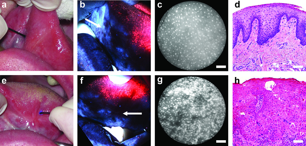
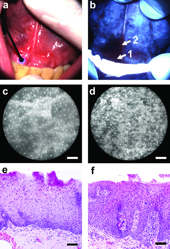
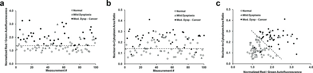
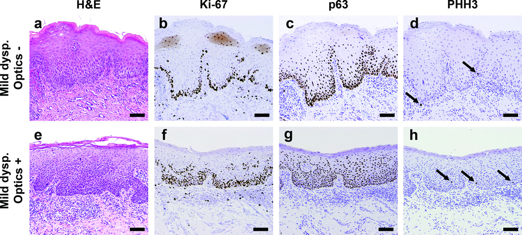
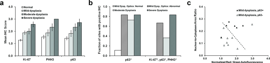
If detected early, oral cancer is eminently curable. However, survival rates for oral cancer patients remain low, largely due to late-stage diagnosis and subsequent difficulty of treatment. To improve clinicians' ability to detect early disease and to treat advanced cancers, we developed a multimodal optical imaging system (MMIS) to evaluate tissue in situ, at macroscopic and microscopic scales. The MMIS was used to measure 100 anatomic sites in 30 patients, correctly classifying 98% of pathologically confirmed normal tissue sites, and 95% of sites graded as moderate dysplasia, severe dysplasia, or cancer. When used alone, MMIS classification accuracy was 35% for sites determined by pathology as mild dysplasia. However, MMIS measurements correlated with expression of candidate molecular markers in 87% of sites with mild dysplasia. These findings support the ability of noninvasive multimodal optical imaging to accurately identify neoplastic tissue and premalignant lesions. This in turn may have considerable impact on detection and treatment of patients with oral cancer and other epithelial malignancies.


Conflict of interest statement

Potential conflict(s) of interest / Disclosure:

Dr. Richards-Kortum serves as an unpaid scientific advisor to Remicalm LLC, holds patents related to optical diagnostic technologies that have been licensed to Remicalm LLC, and holds minority ownership in Remicalm LLC. Dr. Gillenwater has a minority equity interest in Onconome, Inc., and serves as an unpaid scientific advisor to Remicalm LLC. Dr. Schwarz holds a patent related to optical diagnostic technologies that has been licensed to Remicalm LLC.

Figures

Figure 1
Figure 1
Multi-modal optical imaging in the oral cavity. (a-d) A normal site on the anterior tongue. (e-h) A site diagnosed with severe dysplasia on the left mid tongue in the same patient. (a,e) White light examination, (b,f) autofluorescence imaging, (c,g) high-resolution microendoscopy, and (d,h) histopathology sections. In (e), scar tissue is apparent, with the smaller lesion alongside. In (b) and (f), arrows indicate the location of the microendoscope probe, as seen in (a) and (e). Scale bars represent 100 µm.
Figure 2
Figure 2
Multi-modal optical imaging at the floor-of-mouth. (a) The entire area appeared normal on clinical examination. (b) Autofluorescence imaging revealed a region with distinct loss of fluorescence intensity at the patient’s right side. (c,d) High-resolution microendoscope images with the probe placed at sites “1” and “2” in panel (b), respectively. (e,f) Histopathology sections from sites “1” and “2” in panel (b). Scale bars represent 100 µm.
Figure 3
Figure 3
Quantification of AFI and HRME images. Symbols represent the diagnosis for each site according to pathology. (a) Normalized ratio of red-to-green autofluorescence intensity at each of the 100 sites measured in the study. (b) Nuclear-to-cytoplasm area ratio for the same 100 sites shown in (a). (c) Classification of measurement sites using both wide-field autofluorescence and high-resolution morphology. Dashed lines represent linear threshold values to discriminate between normal sites, and those with mild / moderate / severe dysplasia or cancer.
Figure 4
Figure 4
Histopathology sections of tissue from two sites with a diagnosis of mild dysplasia. (a-d) Sections from a site which was classified as “normal” by optical measurement. (e-h) Sections from a site classified as “abnormal” by optical measurement. (a,e) H&E stained sections used by the study pathologist to classify each site as mild dysplasia. Immunostained sections from the same sites with (b) Ki-67, graded low, (c) p63, low (d) PHH3, low, (f) Ki-67, moderate, (g) p63, high, (h) PHH3, high. Scale bars represent 100 µm.
Figure 5
Figure 5
Analysis of IHC staining of sections from tissue sites with a pathology diagnosis of normal or dysplasia. (a) Mean IHC score (see main text for details) versus pathology grade established by H&E staining, for each of the markers Ki-67, PHH3, and p63. Error bars represent standard errors. (b) The fraction of tissue sites with a positive IHC score for either p63 alone, or for the complete panel of markers tested (see text for definition). Data are shown separated by the pathology grade established by H&E staining. (c) Plot of wide-field autofluorescence and high-resolution N/C ratio values for sites with a pathology diagnosis of mild dysplasia, stratified by p63 status. Note that 15 out of the original 17 measurement sites are displayed due to IHC processing artifacts in 2 cases. The dashed line represents the linear threshold shown in Fig. 5 to discriminate between normal sites, and those with mild / moderate / severe dysplasia or cancer.

Similar articles

Cited by

References

    1. Ferlay J, Shin HR, Bray F, Forman D, Mathers C, Parkin DM. Cancer Incidence and Mortality Worldwide: IARC CancerBase No. 10. Lyon, France: International Agency for Research on Cancer; 2010. GLOBOCAN 2008 v1.2.
    1. Chen AY, Myers JN. Cancer of the oral cavity. Dis Mon. 2001;47:275–361. - PubMed
    1. Lingen MW, Kalmar JR, Karrison T, Speight PM. Critical evaluation of diagnostic aids for the detection of oral cancer. Oral Oncol. 2008;44:10–22. - PMC - PubMed
    1. Thomson PJ. Field change and oral cancer: new evidence for widespread carcinogenesis? Int J Oral Maxillofac Surg. 2002;31:262–266. - PubMed
    1. De Veld DC, Witjes MJ, Sterenborg HJ, Roodenburg JL. The status of in vivo autofluorescence spectroscopy and imaging for oral oncology. Oral Oncol. 2005;41:117–131. - PubMed

Publication types