Case Reports
. 2012 May-Jun;31(3):127-34.
doi: 10.5414/np300441.

Creutzfeldt-Jakob disease with unusually extensive neuropathology in a child treated with native human growth hormone

Affiliations
Case Reports

Creutzfeldt-Jakob disease with unusually extensive neuropathology in a child treated with native human growth hormone

Jacqueline Mikol et al. Clin Neuropathol. 2012 May-Jun.

Abstract

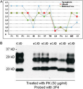
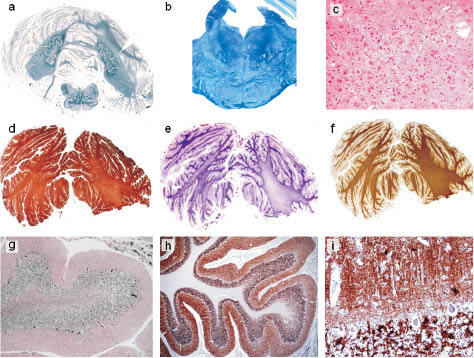
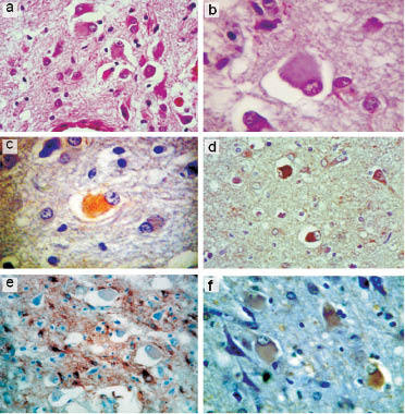
We report a case of iatrogenic Creutzfeldt-Jakob disease(iCJD) in a child with a neonatal growth hormone (GH) deficiency that was treated with native human growth hormone (hGH) between the ages of 9 months and 7 years. Three years after the end of treatment a progressive neurological syndrome consistent with Creutzfeldt-Jakob disease (CJD) developed, leading to death within a year, at age 11. Neuropathological examination showed an unusual widespread form of CJD, notably characterized by (i) involvement of the cerebellar white matter, (ii) cortico-spinal degeneration and (iii) ballooned neurons. A transitional form of the disease between common iatrogenic and panencephalopathic CJD is suggested.


Figures

Figure 1
Figure 1. A: Gray matter lesion profiles: frontal (FC), temporal (TE), parietal (PC) and occipital (OC) neocortices, hippocampus (HI), entorhinal cortex (EC), striatum (caudatus and putamen nuclei) (ST), thalamus (TH), locus niger (LN), midbrain periventricular gray (PG), locus coeruleus (LC), medulla (ME), cerebellum (CB). B: Western blot analysis of PrP. Three sCJD with PrP Type 1 (MM1: 3 lanes at left) and two sCJD with PrP Type 2 (VV2: lanes at right) were used as controls.
Figure 2
Figure 2. a, b, c: View of the left rostral frontal lobe showing cortical atrophy, dilatated ventricle and mild pallor of the white matter in Heidenhain stain (Hd). a: but less in immunostain for MBP (b) prominent PrP deposition in the frontal cortex (c). d: Atrophy of frontal gyri, hematoxylin-eosin (HE) stain × 2. e: Status spongiosus of the frontal cortex, HE stain × 25. f: Low- power view of hemispheric section showing atrophy of cortical ribbon and mild pallor of the white matter in Hd stain. g: Gliosis of frontal WM: immunostain for GFAP × 1.25. h: Massive neuronal loss and gliosis of putamen, HE stain × 62.5. i: Synaptic type PrP deposits in frontal cortex: immunostain with 3F4 antibody × 20.
Figure 3
Figure 3. a: Massive atrophy of the cerebellum with white matter myelin loss. Degeneration of the cortico-spinal tracts, Hd stain × 0.5. b: Pons: degeneration of the cortico-spinal tracts, Luxol fast blue × 2. c: Detail of pons showing neuronal loss and astrocytosis of pontine nuclei: HE stain × 92. d: Gliosis of the lamellae and the album of the cerebellum: immunostain for GFAP. e: Comparison of the Kantzler stain for fibrillary gliosis and f: immunostain for MBP. g: Atrophic cerebellar folia, massive neuronal loss of granule cells, less pronounced of the Purkinje cells, and spongiosis, Bodian impregnation × 12. PrP deposits in the cerebellar cortex of synaptic type with a few plaque-like structures in h: × 2 and i: × 20.
Figure 4
Figure 4. Ballooned neurons with excentric nucleus. a, b, c: cingular cortex, HE stain (a and b) and moderate positivity of the synaptophysin immunostain (c). d: Insular cortex: strong positivity with anti-phosphorylated neurofilament SMI 31. e: Cingular cortex: negative ballooned neurons are surrounded by astrocytes, immunostain for GFAP. f: Weak positivity of BN: immunostain for ubiquitin. a, d and e × 62.5; b, c, d × 125.

Similar articles

Cited by

  • Recombinant human prion protein inhibits prion propagation in vitro.
    Yuan J, Zhan YA, Abskharon R, Xiao X, Martinez MC, Zhou X, Kneale G, Mikol J, Lehmann S, Surewicz WK, Castilla J, Steyaert J, Zhang S, Kong Q, Petersen RB, Wohlkonig A, Zou WQ. Yuan J, et al. Sci Rep. 2013 Oct 9;3:2911. doi: 10.1038/srep02911. Sci Rep. 2013. PMID: 24105336 Free PMC article.

References

    1. Brown P Preece M Brandel JP Sato T McShane L Zerr I Fletcher A Will RG Pocchiari M Cashman NR d’Aignaux JH Cervenáková L Fradkin J Schonberger LB Collins SJ Iatrogenic Creutzfeldt-Jakob disease at the millennium. Neurology. 2000; 55: 1075–1081 - PubMed
    1. INVSMaladie de Creutzfeldt-Jakob et formes apparentées. 2010;
    1. Deslys JP Lasmézas C Dormont D Selection of specific strains in iatrogenic Creutzfeldt-Jakob disease. Lancet. 1994; 343: 848–849 doi:10.1016/S0140-6736(94)92046-X - PubMed
    1. Yuan J Xiao X Qing L Mikol J Ironside JW Gambetti P Kong Q 2009;
    1. Mikol J Goutieres F Deslys JP Parchi P Growth hormone induced Creutzfeldt-Jakob disease (CJD) in a child with extensive lesions. Brain Pathol. 1994; 4:

Publication types

Substances