. 2012 Nov;42(5):878-84.
doi: 10.1093/ejcts/ezs128. Epub 2012 May 2.

Inhibition of cell surface expression of endothelial adhesion molecules by ursolic acid prevents intimal hyperplasia of venous bypass grafts in rats

Affiliations

Inhibition of cell surface expression of endothelial adhesion molecules by ursolic acid prevents intimal hyperplasia of venous bypass grafts in rats

Iris Zeller et al. Eur J Cardiothorac Surg. 2012 Nov.

Abstract

Objectives: Despite rapid progress in surgical techniques, there is still a significant lack of surgery-supportive pharmacological treatments. The aim of this study was to test the hypothesis that ursolic acid (UA) may prevent intimal hyperplasia of venous bypass grafts.

Methods: The hypothesis was tested by means of primary cell isolation and culture followed by real-time polymerase chain reaction, western blotting, fluorescence microscopy and fluorescence-activated cell sorting analyses, as well as an in vivo rat model for intimal hyperplasia of venous bypass grafts and immunohistochemistry and histochemistry.

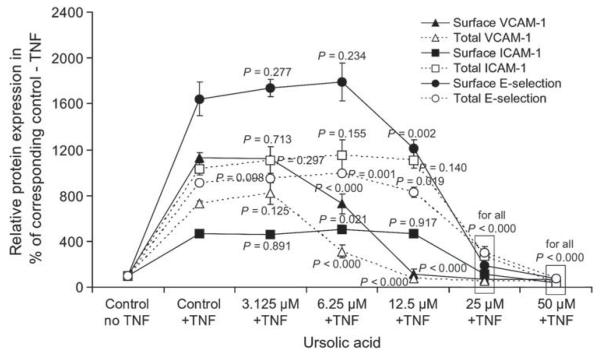
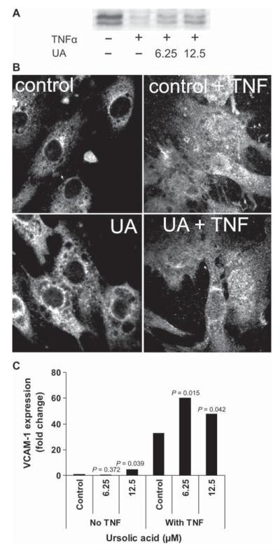
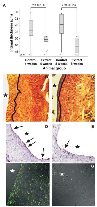
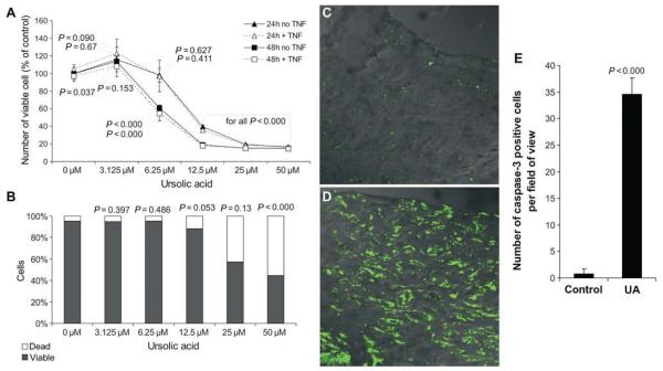
Results: The local application of UA significantly inhibited intimal hyperplasia in vivo (intimal thickness control: 25 µm, UA group: 18 µM-8 weeks after surgery). The UA treatment of grafts significantly resulted in reduced endothelial vascular cell adhesion molecule-1 (VCAM-1) expression, reduced infiltration of the grafts vessel wall by CD45-positive cells and increased smooth muscle cell (SMC) death. In in vitro condition, it could be shown that UA inhibits VCAM-1 expression downstream of NFκB and is likely to interfere with VCAM-1 protein synthesis in endothelial cells. Quantification of cell death in vascular smooth muscle cells treated with UA indicated that UA is a potent inducer of SMC apoptosis.

Conclusions: Our results suggest that UA-mediated inhibition of endothelial VCAM-1 expression reduces the infiltration of venous bypass grafts by CD45-positive cells and inhibits intimal hyperplasia. Apoptosis induction in SMCs may be another method in which UA reduces intimal thickening. UA may constitute a surgery-supportive pharmacon that reduces intimal hyperplasia of vein grafts.


Figures

Figure 1
Figure 1
UA inhibits TNFα-mediated VCAM-1, ICAM-1 and E-selectin protein expression in HUVECs. To test whether UA has an effect on TNFα-induced expression of adhesion molecules (VCAM-1, ICAM-1 and E-selectin), we analysed protein expression (surface and total) in a previously developed ELISA-based system. HUVECs were treated with UA (final concentrations: 3.125, 6.25, 12.5, 25 and 50 μM) or DMSO (as solvent control) and 100 ng/ml of TNFα was indicated. Incubation times were 4 (E-selectin) or 15 h (VCAM-1 and ICAM-1). Mean ± SD values of a representative experiment performed in quadruplicate are shown. The experiment was repeated three times, giving similar results.
Figure 2
Figure 2
UA prevents TNFα-mediated degradation of IκB, but does not inhibit NFκB translocation and function in HUVECs. (A) The western blot analysis of IκB. Cells were treated with DMSO (as a solvent control), 6.25 and 12.5 μM UA 30 min before the addition of 100 ng/ml of TNFα. Samples were taken 30 min after the addition of TNFα. A representative blot is shown. (B) An immunofluorescence analysis of NFκB (p65). Cells were treated with DMSO (as a solvent control) or 12.5 μM UA 30 min before the addition of 100 ng/ml of TNFα, and fixed 30 min thereafter. Representative images are shown. (C) The real-time PCR data of VCAM-1 expression in HUVECs. The cells were treated with DMSO (as a solvent control), 6.25 and 12.5 μM UA 30 min before the addition of 100 ng/ml of TNFα. Cells were harvested 6 h after the addition of TNFα. Data shown are mean values of three experiments performed in duplicates. All experiments were repeated at least three times.
Figure 3
Figure 3
Sambucus ebulus diethyl ether extract is a potent inhibitor of vein graft intimal hyperplasia in vivo. (A) The boxplot summarizes the data on the effects of UA (S. ebulus diethyl ether) on intimal thickening compared with the control, 4 and 8 weeks after grafting (n per group = 10). Six sections per graft were analysed by blinded researchers. (B:’ control) and (C: S. ebulus extract) show an Elastica van Giesson staining (dark lines indicate the intima-media-border), (D: control) and (E: S. ebulus extract) show immunostaining for VCAM-1 (arrows), and (F: control) and (G: S. ebulus) extract show CD45-positive cell infiltrates in graft tissue sections (×100) collected at 8 weeks post grafting. Representative images are shown. The asterisks indicate the vessel lumen.
Figure 4
Figure 4
UA inhibits SMC proliferation and induces SMC cell death in vitro and in vivo. Cells were treated with 3.125, 6.25, 12.5, 25 and 50 μM UA or DMSO (solvent control) and subjected to an analysis of the number of viable cells (A) and cell death analysis (B - 24 h incubation). Data shown are mean values ± SD of representative experiments performed in triplicates. (C: control) and (D: S. ebulus) extract show an immunofluorescence staining for active caspase-3 within the intima on sections of the animal experiment (×400); time point: 8 weeks. Representative images are shown. (E) The boxplot shows the quantification of cells positive for active caspase-3. Four fields of view were analysed per sample.

Similar articles

Cited by

References

    1. Crook MF, Newby AC, Southgate KM. Expression of intercellular adhesion molecules in human saphenous veins: effects of inflammatory cytokines and neointima formation in culture. Atherosclerosis. 2000;150:33–41. - PubMed
    1. Hansson GK. Inflammatory mechanisms in atherosclerosis. J Thromb Haemost. 2009;7:328–31. - PubMed
    1. Lau GT, Ridley LJ, Bannon PG, Wong LA, Trieu J, Brieger DB, et al. Lumen loss in the first year in saphenous vein grafts is predominantly a result of negative remodeling of the whole vessel rather than a result of changes in wall thickness. Circulation. 2006;114:1435–140. - PubMed
    1. Blankenberg S, Barbaux S, Tiret L. Adhesion molecules and atherosclerosis. Atherosclerosis. 2003;170:191–203. - PubMed
    1. Chi Z, Melendez AJ. Role of cell adhesion molecules and immune-cell migration in the initiation, onset and development of atherosclerosis. Cell Adhes Migr. 2007;1:171–5. - PMC - PubMed

Publication types

MeSH terms