. 2012 Jul;132(7):1860-8.
doi: 10.1038/jid.2011.451. Epub 2012 May 3.

Histology-specific microRNA alterations in melanoma

Affiliations

Histology-specific microRNA alterations in melanoma

Laura Poliseno et al. J Invest Dermatol. 2012 Jul.

Abstract

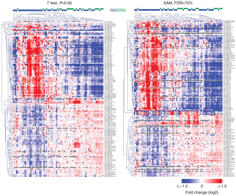
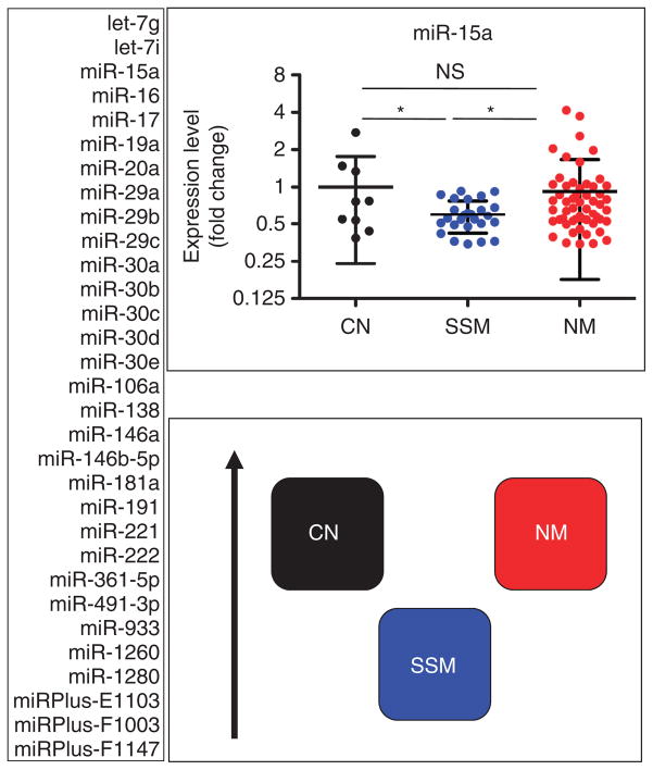
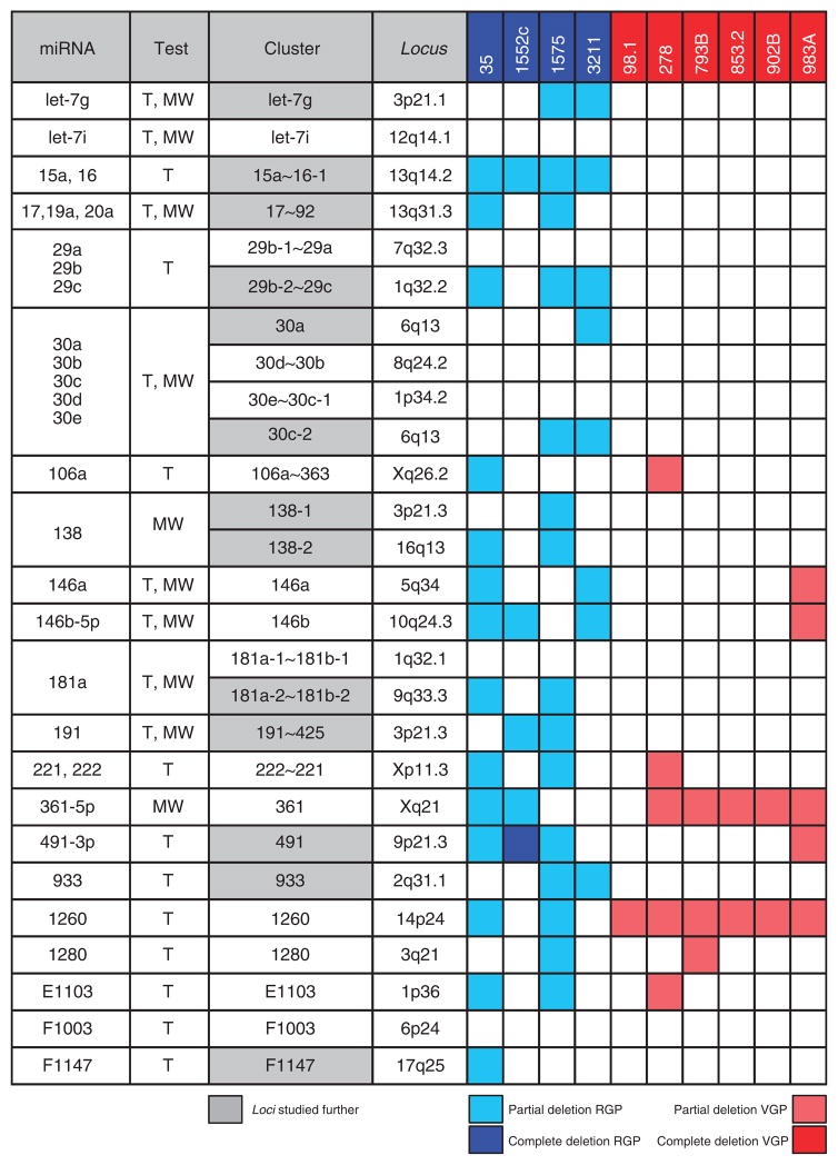
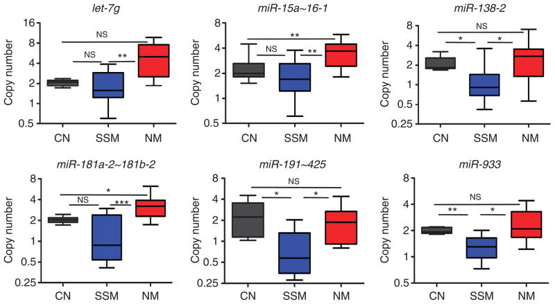
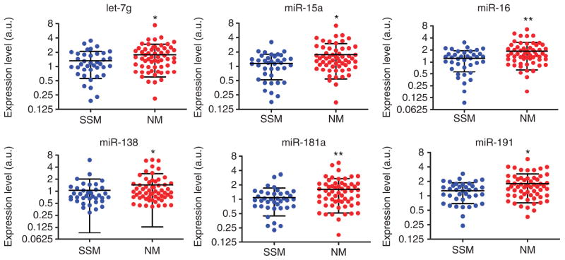
We examined the microRNA signature that distinguishes the most common melanoma histological subtypes, superficial spreading melanoma (SSM) and nodular melanoma (NM). We also investigated the mechanisms underlying the differential expression of histology-specific microRNAs. MicroRNA array performed on a training cohort of 82 primary melanoma tumors (26 SSM, 56 NM), and nine congenital nevi (CN) revealed 134 microRNAs differentially expressed between SSM and NM (P<0.05). Out of 134 microRNAs, 126 remained significant after controlling for thickness and 31 were expressed at a lower level in SSM compared with both NM and CN. For seven microRNAs (let-7g, miR-15a, miR-16, miR-138, miR-181a, miR-191, and miR-933), the downregulation was associated with selective genomic loss in SSM cell lines and primary tumors, but not in NM cell lines and primary tumors. The lower expression level of six out of seven microRNAs in SSM compared with NM was confirmed by real-time PCR on a subset of cases in the training cohort and validated in an independent cohort of 97 melanoma cases (38 SSM, 59 NM). Our data support a molecular classification in which SSM and NM are two molecularly distinct phenotypes. Therapeutic strategies that take into account subtype-specific alterations might improve the outcome of melanoma patients.


Conflict of interest statement

CONFLICT OF INTEREST

The authors declare no conflict of interest.

Figures

Figure 1
Figure 1. Expression profiling reveals 134 microRNAs that are differentially expressed between superficial spreading melanoma (SSM) and nodular melanoma (NM)
Heat map representing the differentially expressed microRNAs according to T-test, P<0.05 (left), and SAM, false discovery rate (FDR) <10% (right). T-test and SAM identify 160 and 165 differentially expressed microRNAs, respectively. In all, 134 microRNAs overlap between the two analyses. For 126 of the 134 microRNAs, the differences between SSM and NM could not be accounted for by thickness.
Figure 2
Figure 2. MicroRNAs that are specifically downregulated in superficial spreading melanoma (SSM) compared with both congenital nevi (CN) and nodular melanoma (NM)
In all, 31 microRNAs belong to this category according to T-test and/or Mann–Whitney test (P<0.05), and they are all listed on the left. On the right, miR-15a is shown as an example in the upper panel, whereas a cartoon summarizing the relationship among CN, SSM, and NM is reported in the lower panel. The black arrow on the side indicates increasing expression levels. *P<0.05.
Figure 3
Figure 3. Thirteen microRNA loci are deleted in radial growth phase (RGP)/superficial spreading melanoma (SSM)–like cell lines and not in vertical growth phase (VGP)/nodular melanoma (NM)–like cell lines
The genomic loci corresponding to the 31 microRNAs listed in Figure 2 were analyzed by real-time PCR in 10 primary melanoma cell lines. The second column indicates whether each microRNA was identified using T-test (T) or Mann–Whitney test (MW). The cluster to which each microRNA belongs and its genomic location are reported in the third and fourth column, respectively. RGP/SSM-like cell lines are in blue and VGP/NM-like cell lines are in red. The 13 loci that were subjected to further analyses are highlighted in gray.
Figure 4
Figure 4. Validation of the genomic loss of let-7g, miR-15a, miR-16, miR-138, miR-181a, miR-191, and miR-933 in melanoma specimens
Copy number of the microRNA loci corresponding to the seven microRNAs in congenital nevi (CN) (black), superficial spreading melanoma (SSM) (blue), and nodular melanoma (NM) (red) primary tumor specimens. The bicistronic miR-15a~16-1 expresses both miR-15a and miR-16. *P<0.05; **P<0.01; ***P<0.001.
Figure 5
Figure 5. Expression level of let-7g, miR-15a, miR-16, miR-138, miR-181a, and miR-191 in the melanoma specimens belonging to the validation cohort
The difference in expression level of the indicated microRNAs between the 38 superficial spreading melanoma (SSM) and the 59 nodular melanoma (NM) samples belonging to the validation cohort is reported. *P<0.05; **P<0.01.

Similar articles

Cited by

References

    1. Akavia UD, Litvin O, Kim J, et al. An integrated approach to uncover drivers of cancer. Cell. 2010;143:1005–17. - PMC - PubMed
    1. Baillat D, Shiekhattar R. Functional dissection of the human TNRC6 (GW182-related) family of proteins. Mol Cell Biol. 2009;29:4144–55. - PMC - PubMed
    1. Baumert J, Schmidt M, Giehl KA, et al. Time trends in tumour thickness vary in subgroups: analysis of 6475 patients by age, tumour site and melanoma subtype. Melanoma Res. 2009;19:24–30. - PubMed
    1. Calin GA, Sevignani C, Dumitru CD, et al. Human microRNA genes are frequently located at fragile sites and genomic regions involved in cancers. Proc Natl Acad Sci USA. 2004;101:2999–3004. - PMC - PubMed
    1. Caramuta S, Egyhazi S, Rodolfo M, et al. MicroRNA expression profiles associated with mutational status and survival in malignant melanoma. J Invest Dermatol. 2010;130:2062–70. - PubMed

Publication types