. 2012 Jun;37(6):611-20.
doi: 10.1097/SHK.0b013e318254fa6a.

The role and source of tumor necrosis factor-α in hemorrhage-induced priming for septic lung injury

Affiliations

The role and source of tumor necrosis factor-α in hemorrhage-induced priming for septic lung injury

Joanne Lomas-Neira et al. Shock. 2012 Jun.

Abstract

Tumor necrosis factor α (TNF-α) has been reported to be a key component of the functional priming, of both myeloid and nonmyeloid cells, that is thought to contribute to the lung's increased susceptibility to injury following shock. Not surprisingly, we found that mice deficient in TNF-α exhibited reduced acute lung injury (ALI) resultant from the combined insults of hemorrhagic shock and sepsis. However, we found that when we adoptively transferred neutrophils from mice expressing TNF-α to neutrophil-depleted mice that lacked TNF-α, they were not able to serve as priming stimulus for the development of ALI. Based on these findings, we proposed that resident lung tissue cells mediate TNF-α priming. To begin to unravel the complex signaling pathway of various resident lung tissue cells in TNF-α-induced priming, we compared the effect of local (intratracheal [i.t.]) versus systemic [intravenous (i.v.)] delivery of TNF-α small interference (siRNA). We hypothesized that alternately suppressing expression of TNF-α in lung endothelial (i.v.) or epithelial (i.t.) cells would produce a differential effect in shock-induced ALI. We found that when in vivo siRNA i.t. or i.v. against TNF-α was administered to C57/BL6 mice at 2 h after hemorrhage, 24 h before septic challenge, that systemic/i.v., but not i.t., delivery of TNF-α siRNA following hemorrhage priming significantly reduces expression of indices of ALI compared with controls. These findings suggest that an absence of local lung tissue TNF-α significantly reduces lung tissue injury following hemorrhage priming for ALI and that pulmonary endothelial and/or other possible vascular resident cells, not epithelial cells, play a greater role in mediating the TNF-α priming response in a mouse model of hemorrhage/sepsis-induced ALI.


Conflict of interest statement

Authors have no existing conflicts of interest related to the subject matter of this manuscript.

Figures

Figure 1
Figure 1
Murine model of hemorrhage-induced priming for the development of ALI (A). Model for the study of adoptive transfer of hemorrhage-induced priming for the development of ALI (B).
Figure 2
Figure 2
Lung tissue homogenate levels of pro-inflammatory cytokine, IL-6 (A), and neutrophil chemotactic proteins, MIP-2 (D), were significantly decreased in lungs from Hem/CLP TNF−/− mice when compared with Hem/CLP background, TNF+/+ mice. Neutrophil chemotactic protein, KC, was significantly decreased in lung tissue (C) and IL-10 (F) showed significant decreased in plasma. (n=7–8. *p<0.05 vs. equivalent Sham, @
Figure 3
Figure 3
Plasma levels of IL-6 (A), and neutrophil chemotactic proteins, KC (C) and MIP-2 (D), in blood were significantly decreased following Hem/CLP in TNF−/− mice as compared to background, TNF+/+ mice. This was not observe in plasma IL-10 levels (B), where only mice in the Sham Hem (SHem)/CLP groups showed significant difference.
Figure 4
Figure 4
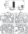
Lung tissue myeloperoxidase activity (A), a measure of neutrophil influx to lung, was reduced greater than 50% in lungs from Hem/CLP, TNF−/− mice relative to Hem/CLP TNF+/+ mice. This is consistent with histological findings of significantly reduced lung tissue septal thickening and cellular infiltrate (E) and esterase+ (neutrophil specific) cells (E/insert box) compared to lung tissue from Hem/CLP TNF+/+ mice (D/insert box). (n=7–8. *p<0.05 vs. equivalent Sham, @
Figure 5
Figure 5
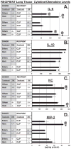
TNF−/− recipients that received neutrophils from Hem TNF+/+ donors showed significantly reduced levels of IL-6 (A) in lung tissue homogenates in contrast to TNF+/+ competent Donor/Recipient and TNF+/+ Recipients that received TNF−/− neutrophils from Hem Donors. However, IL-10 (B), while reduced between Donor treatments (SHem vs. Hem) was not different between neutrophil Donor/lung tissue Recipient combinations. Levels of KC (C) and MIP-2 (D) in TNF−/− Recipients were significantly reduced in contrast to TNF+/+ Donor/Recipient combination, however, TNF−/− Donor neutrophils also reduced MIP-2 in TNF+/+ recipients (D). (n=7–8. *p<0.05 vs. equivalent Sham, @
Figure 6
Figure 6
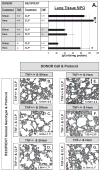
Lung tissue myeloperoxidase activity (A), was significantly reduced in TNF−/− recipient lung tissue compared to either TNF+/+ Recipient combination (n=7–8. *p<0.05 vs. equivalent Sham, @
Figure 7
Figure 7
Confocal microscopy of i.v. delivered, fluorochrome-labeled (SiGlo RISC-free) siRNA uptake in lung endothelial (Tie2+) cells (A). Uptake of Alexa 647 labeled endothelial cell growth factor, Angiopoietin-2, siRNA via i.v. delivery was observed in CD31+ (endothelial cells), while i.t. delivered siRNA was observed in cytokeratin 18+ (epithelial cells) and a small percent of macrophages (as assessed by forward/side scatter analysis) (B).
Figure 8
Figure 8
i.v. delivery of TNF-α siRNA 2 hours post hemorrhage priming significantly reduced levels of TNF-α (C), IL-6 (E) as well as MPO activity (G) in mouse lungs following CLP when compared with siRNA control, this was not observed in the naked i.t. delivered TNF-α treated mice. (n=6–7. * p< 0.05 vs. SHem/CLP control, #p<0.05 vs. Hem/CLP Control-siRNA). TNF-α gene expression in lung tissue homogenates, as measured by total RNA (Rt-PCR) (n=4), was consistently lower following i.v. as opposed to i.t delivery of TNF-α siRNA (8A).
Figure 9
Figure 9
Comparison of representative hemotoxylin and eosin stained lung tissue sections show a discernible decrease in lung inflammation, cellular infiltrate and septal thickening, in the i.v. TNF-α siRNA (D), but not i.t. (C) TNF-α siRNA route of delivery when compared to Hem/CLP control mice (B). (n=4/group)

Similar articles

Cited by

References

    1. Abraham E, Carmody A, Shenkar R, Arcaroli J. Neutrophils as early immunologic effectors in hemorrhage- or endotoxemia-induced acute lung injury. Am J Physiol Lung Cell Mol Physiol. 2000;279:L1137–L1145. - PubMed
    1. Alarcon P, Conejeros I, Carretta MD, Concha C, Jara E, Tadich N, Hidalgo MA, Burgos RA. D-Lactic acid interferes with the effects of platelet activating factor on bovine neutrophils. Veterinary Immun and Immunopath. 2011;144:68–78. - PubMed
    1. Aleku M, Fisch G, Mopert K, Keil O, Woflgang A, Kaufmann J, Santel A. Intracellular localization of lipoplexed siRNA in vascular endothelial cells of different mouse tissues. Microvasc Res. 2008;76:31–41. - PubMed
    1. Angus DC, Linde-Zwirble WT, Lidicker J, Clermont G, Carcillo J, Pinsky MR. Epidemiology of severe sepsis in the United States: analysis of incidence, outcome, and associated costs of care. Crit Care Med. 2001;29:1303–1310. - PubMed
    1. Ayala A, Chung CS, Lomas J, Grutkoski PS, Doughty LA, Simms HH. A mouse model of priming for acute lung injury following shock. Shock. 2001;15:83S.

Publication types

Substances