

High-strength silk protein scaffolds for bone repair
- PMID: 22552231
- PMCID: PMC3356671
- DOI: 10.1073/pnas.1119474109
High-strength silk protein scaffolds for bone repair
Abstract
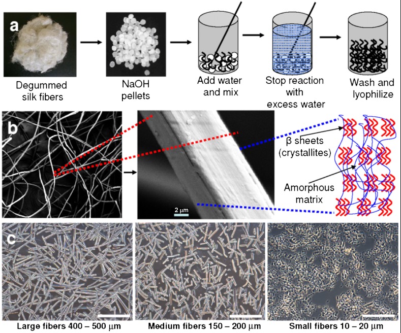
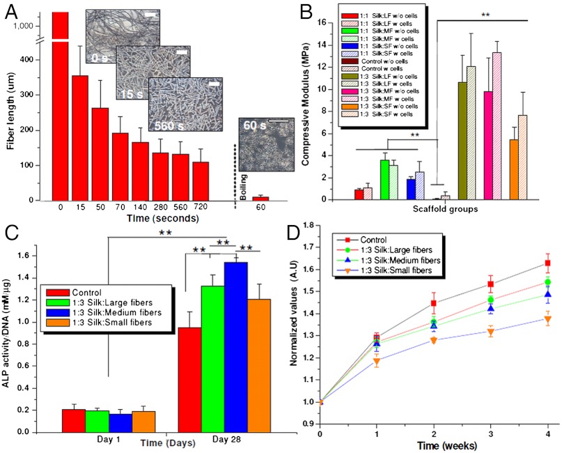
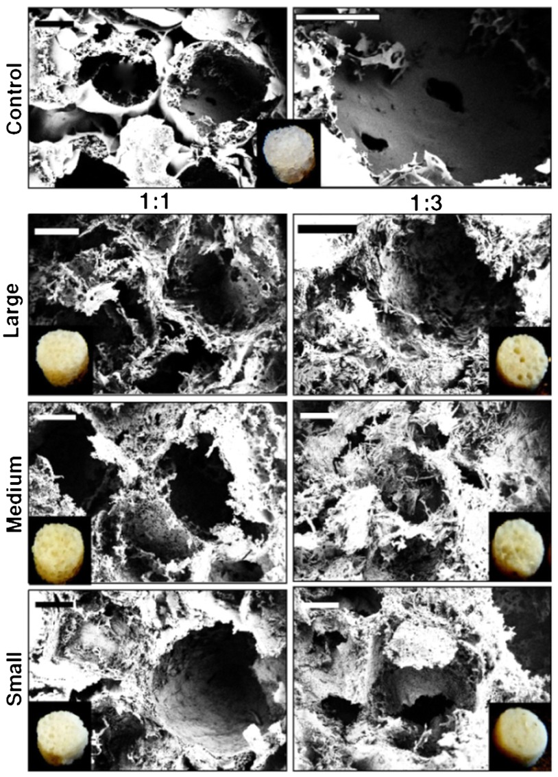
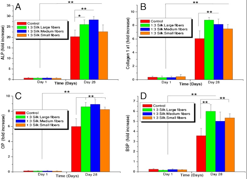
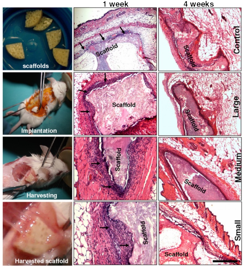
Biomaterials for bone tissue regeneration represent a major focus of orthopedic research. However, only a handful of polymeric biomaterials are utilized today because of their failure to address critical issues like compressive strength for load-bearing bone grafts. In this study development of a high compressive strength (~13 MPa hydrated state) polymeric bone composite materials is reported, based on silk protein-protein interfacial bonding. Micron-sized silk fibers (10-600 µm) obtained utilizing alkali hydrolysis were used as reinforcement in a compact fiber composite with tunable compressive strength, surface roughness, and porosity based on the fiber length included. A combination of surface roughness, porosity, and scaffold stiffness favored human bone marrow-derived mesenchymal stem cell differentiation toward bone-like tissue in vitro based on biochemical and gene expression for bone markers. Further, minimal in vivo immunomodulatory responses suggested compatibility of the fabricated silk-fiber-reinforced composite matrices for bone engineering applications.
Conflict of interest statement
The authors declare no conflict of interest.
Figures
References
-
- Drosse I. Tissue engineering for bone defect healing: An update on a multi-component approach. Injury. 2008;39(Suppl 2):S9–S20. - PubMed
-
- Langer R, Vacanti JP. Tissue engineering. Science. 1993;260:920–926. - PubMed
-
- Marquis ME, et al. Bone cells biomaterials interactions. Front Biosci. 2009;14:1023–1067. - PubMed
-
- Khan Y, Yaszemski MJ, Mikos AG, Laurencin CT. Tissue engineering of bone: Material and matrix considerations. J Bone Joint Surg Am. 2008;90:36–42. - PubMed
-
- Dawson JI, et al. Development of specific collagen scaffolds to support the osteogenic and chondrogenic differentiation of human bone marrow stromal cells. Biomaterials. 2008;29:3105–3116. - PubMed
Publication types
MeSH terms
Substances
Grants and funding
LinkOut - more resources
Full Text Sources
Other Literature Sources
