. 2012 Jul 1;72(13):3260-9.
doi: 10.1158/0008-5472.CAN-11-4141. Epub 2012 May 2.

Oncogenic PI3K mutations lead to NF-κB-dependent cytokine expression following growth factor deprivation

Affiliations

Oncogenic PI3K mutations lead to NF-κB-dependent cytokine expression following growth factor deprivation

Jessica E Hutti et al. Cancer Res. .

Abstract

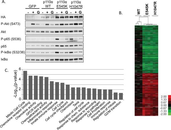
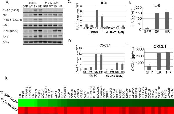
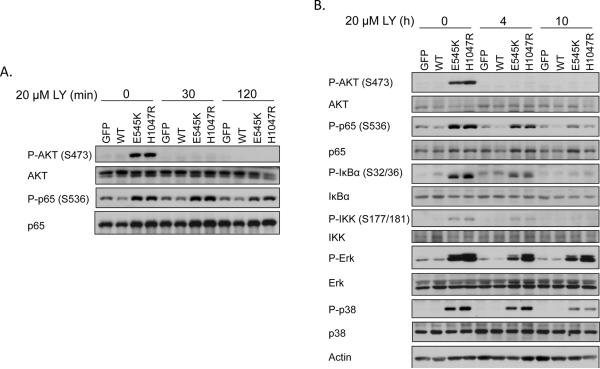
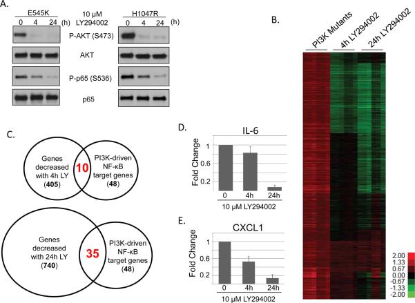
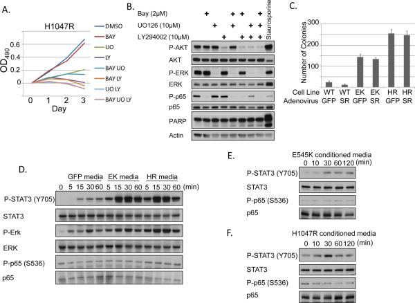
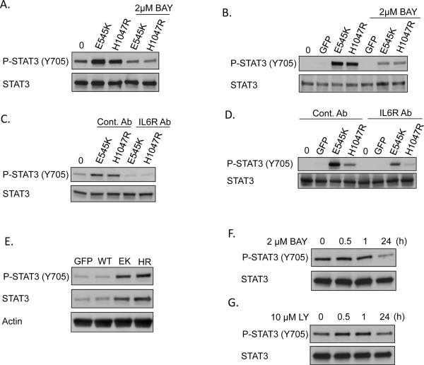
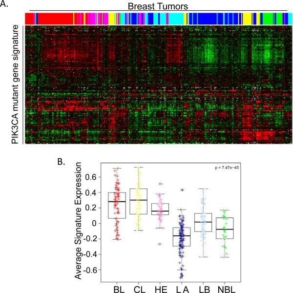
The phosphoinositide 3-kinase (PI3K) pathway is one of the most commonly misregulated signaling pathways in human cancers, but its impact on the tumor microenvironment has not been considered as deeply as its autonomous impact on tumor cells. In this study, we show that NF-κB is activated by the two most common PI3K mutations, PIK3CA E545K and H1047R. We found that markers of NF-κB are most strongly upregulated under conditions of growth factor deprivation. Gene expression analysis conducted on cells deprived of growth factors identified the repertoire of genes altered by oncogenic PI3K mutations following growth factor deprivation. This gene set most closely correlated with gene signatures from claudin-low and basal-like breast tumors, subtypes frequently exhibiting constitutive PI3K/Akt activity. An NF-κB-dependent subset of genes driven by oncogenic PI3K mutations was also identified that encoded primarily secreted proteins, suggesting a paracrine role for this gene set. Interestingly, while NF-κB activated by oncogenes such as Ras and EGF receptor leads to cell-autonomous effects, abrogating NF-κB in PI3K-transformed cells did not decrease proliferation or induce apoptosis. However, conditioned media from PI3K mutant-expressing cells led to increased STAT3 activation in recipient THP-1 monocytes or normal epithelial cells in a NF-κB and interleukin-6-dependent manner. Together, our findings describe a PI3K-driven, NF-κB-dependent transcriptional profile that may play a critical role in promoting a microenvironment amenable to tumor progression. These data also indicate that NF-κB plays diverse roles downstream from different oncogenic signaling pathways.


Figures

Figure 1
Figure 1. IKK/NF-κB is activated by oncogenic PI3K mutations
MCF10A cells stably expressing HA-GFP, HA-PIK3CA WT, HA-PIK3CA E545K, or HA-PIK3CA H1047R were generated by retroviral transduction. A) Cells were grown in growth medium (G), starvation medium lacking EGF and insulin (−) for 24h, or were starved for 24h and stimulated for 10 minutes with 20ng/mL EGF and 10μg/mL insulin (+). Lysates were evaluated by immunoblot. B) Cells expressing PIK3CA WT, E545K, or H1047R were GF-deprived for 24h. RNAs were evaluated by microarray and compared to expression in GFP control samples. Genes which are statistically significant by SAM analysis and altered more than 2-fold are shown in the heat map. Color key is for log2 ratio. C) A categorical enrichment analysis was performed on the top 300 genes upregulated by the E545K or H1047R mutation. Statistically enriched categories are shown (P < 0.05).
Figure 2
Figure 2. NF-κB target genes are upregulated by oncogenic PI3K mutations
MCF10A cells stably expressing GFP, PIK3CA WT, E545K, or H1047R were starved for 24h and treated with DMSO or 2μM BAY-65-1942 for 4h. A) Cells were lysed and evaluated by immunoblot. B) Microarray analysis was performed. Genes which exhibit both increased expression in PIK3CA-transformed cells and decreased expression following IKK inhibition are shown. C–D) Real-time RT-PCR was used to verify expression changes for IL-6 and CXCL1. E–F) ELISA analysis shows increased secretion of IL-6 and CXCL1 by cells expressing the oncogenic PI3K mutations following 24h GF-deprivation.
Figure 3
Figure 3. Sustained PI3K inhibition decreases NF-κB activation by oncogenic PI3K mutations
A) MCF10A cells stably expressing GFP, PIK3CA WT, E545K, or H1047R were starved for 24h, treated with 20μM LY294002 for 0–120 minutes and lysates were evaluated by immunoblot. B) MCF10A cells stably expressing GFP, PIK3CA WT, E545K, or H1047R were starved for 24h, treated with 20μM LY294002 for 0–10h and lysates were evaluated by immunoblot for the indicated signaling molecules.
Figure 4
Figure 4. Sustained PI3K inhibition decreases expression of NF-κB target genes in cells expressing oncogenic PI3K mutations
A) MCF10A cells expressing PIK3CA H1047R were treated for 4h or 24h with 10μM LY294002 and analyzed by immunoblot. B) RNA samples from cells treated as in A) were evaluated by microarray. C) Venn diagram depicting changes in NF-κB-dependent gene expression following 4h or 24h of LY294002 treatment. C–D) MCF10A cells expressing PIK3CA E545K were treated for 4h or 24h with 10μM LY294002 and analyzed by real-time RT-PCR to evaluate expression of C) IL-6 or D) CXCL1.
Figure 5
Figure 5. Secreted factors from cells expressing oncogenic PI3K mutations activate STAT3
A) GF-deprived MCF10A cells expressing PIK3CA H1047R were treated for 3 days with the indicated inhibitors. Media and inhibitors were replaced daily. BAY = 2μM BAY-65-1942. LY = 10μM LY294002. UO = 10μM UO126 and cells were evaluated by MTT assay. B) Growth factor-starved MCF10A cells expressing H1047R were treated for 2 days with the indicated inhibitors. Media and inhibitors were replaced daily. Lysates were evaluated for PARP cleavage. 1μM staurosporine (positive control) was added 6h prior to lysis. C) MCF10A cells expressing PIK3CA WT, E545K, or H1047R were infected with adenovirus expressing either GFP or IκBα superrepressor (SR) and plated in 0.6% Bacto agar in MCF10A media lacking EGF and insulin. Colonies were counted after 25 days. D) MCF10A cells expressing GFP, PIK3CA E545K, or PIK3CA H1047R were starved for 24h. Conditioned media from these cells was used to treat THP-1 monocytes for 0–60 minutes. Lysates were evaluated by immunoblot. E–F) MCF10A cells stably expressing PIK3CA E545K (E) or H1047R (F) were starved for 24h. Conditioned media from these cells was then used to stimulate starved parental MCF10A cells for 0–120 minutes. Lysates were evaluated by immunoblot.
Figure 6
Figure 6. IL-6 contributes to STAT3 activation by E545K or H1047R conditioned media
A) MCF10A cells stably expressing PIK3CA E545K or H1047R were starved for 24h in the presence or absence of 2μM BAY-65-1942. Conditioned media was used to stimulate parental MCF10A cells for 30 min and lysates were evaluated by immunoblot. B) MCF10A cells expressing GFP, PIK3CA E545K, or PIK3CA H1047R were starved for 24h in the presence or absence of 2μM BAY-65-1942. Conditioned media was used to stimulate THP-1 cells for 15 minutes and lysates were evaluated by immunoblot. C) Donor MCF10A cells expressing PIK3CA E545K or H1047R were starved for 24h. Both recipient parental MCF10A cells and donor cells were pretreated for 2h with 500 ng/mL IL-6 receptor antagonizing antibody or conrol antibody (anti-Flag mouse monoclonal). Conditioned media from donor cells was used to stimulate parental MCF10A cells for 30 minutes and lysates were evaluated by immunoblot. D) Donor MCF10A cells expressing GFP, PIK3CA E545K, or PIK3CA H1047R were starved for 24h. Recipient THP-1 cells and donor cells were pretreated for 2h with 500 ng/mL IL-6 receptor antagonizing antibody or control antibody (anti-Flag mouse monoclonal). Conditioned media from donor cells was used to stimulate THP-1 cells for 15 minutes and lysates were evaluated by immunoblot. E) MCF-10A cells expressing GFP, or PIK3CA WT, E545K, or H1047R were GF-deprived for 24h and immunoblotted with the indicated antibodies. F–G) MCF10A cells expressing PIK3CA H1047R were GR-deprived for 24h and treated for the indicated times with F) 2μM BAY-65-1942 or G) 10μM LY294002 and immunoblotted with the indicated antibodies.
Figure 7
Figure 7. The PIK3CA mutant gene signature correlates with claudin low and basal breast cancer subtypes
A) 192 of the top 300 genes upregulated by PIK3CA mutation were identified in the UNC337 dataset. Expression of these genes is shown for each of the breast tumor samples present in the UNC337 dataset. Color-coding denotes tumor subtype: basal-like (red); claudin-low (yellow); HER2-enriched (pink); luminal A (dark blue); luminal B (light blue); normal breast-like (green). B) The expression value for each of the 192 PIK3CA upregulated genes was plotted by subtype and average signature value was calculated. Colored dots or boxes denote tumor subtype. BL (red), basal-like; CL (yellow), claudin-low H2 (pink), HER2-enriched; LA (dark blue), luminal A; LB (light blue), luminal B; NBL (green), normal breast-like.

Similar articles

Cited by

References

    1. Cantley LC. The phosphoinositide 3-kinase pathway. Science. 2002;296(5573):1655–7. - PubMed
    1. Luo J, Manning BD, Cantley LC. Targeting the PI3K-Akt pathway in human cancer: rationale and promise. Cancer cell. 2003;4(4):257–62. - PubMed
    1. Engelman JA, Luo J, Cantley LC. The evolution of phosphatidylinositol 3-kinases as regulators of growth and metabolism. Nat Rev Genet. 2006;7(8):606–19. - PubMed
    1. Vivanco I, Sawyers CL. The phosphatidylinositol 3-Kinase AKT pathway in human cancer. Nature reviews Cancer. 2002;2(7):489–501. - PubMed
    1. Hennessy BT, Gonzalez-Angulo AM, Stemke-Hale K, Gilcrease MZ, Krishnamurthy S, Lee JS, et al. Characterization of a naturally occurring breast cancer subset enriched in epithelial-to-mesenchymal transition and stem cell characteristics. Cancer research. 2009;69(10):4116–24. - PMC - PubMed

Publication types