. 2012 Jul 15;72(14):3652-63.
doi: 10.1158/0008-5472.CAN-12-0128. Epub 2012 May 2.

Temporary disruption of the blood-brain barrier by use of ultrasound and microbubbles: safety and efficacy evaluation in rhesus macaques

Affiliations

Temporary disruption of the blood-brain barrier by use of ultrasound and microbubbles: safety and efficacy evaluation in rhesus macaques

Nathan McDannold et al. Cancer Res. .

Abstract

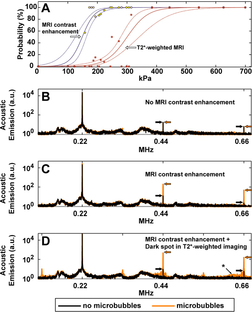
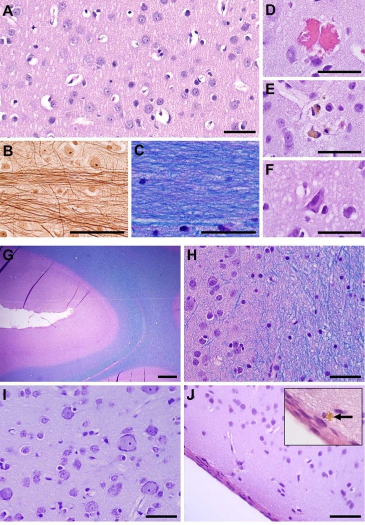
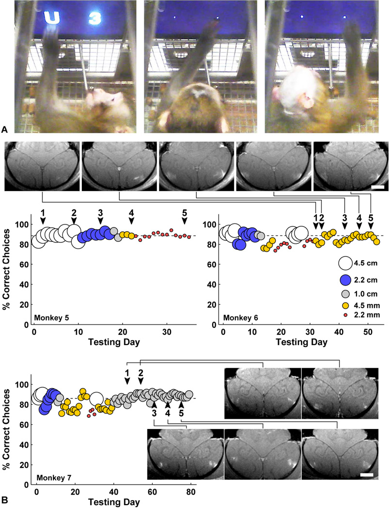
The blood-brain barrier (BBB) prevents entry of most drugs into the brain and is a major hurdle to the use of drugs for brain tumors and other central nervous system disorders. Work in small animals has shown that ultrasound combined with an intravenously circulating microbubble agent can temporarily permeabilize the BBB. Here, we evaluated whether this targeted drug delivery method can be applied safely, reliably, and in a controlled manner on rhesus macaques using a focused ultrasound system. We identified a clear safety window during which BBB disruption could be produced without evident tissue damage, and the acoustic pressure amplitude where the probability for BBB disruption was 50% and was found to be half of the value that would produce tissue damage. Acoustic emission measurements seem promising for predicting BBB disruption and damage. In addition, we conducted repeated BBB disruption to central visual field targets over several weeks in animals trained to conduct complex visual acuity tasks. All animals recovered from each session without behavioral deficits, visual deficits, or loss in visual acuity. Together, our findings show that BBB disruption can be reliably and repeatedly produced without evident histologic or functional damage in a clinically relevant animal model using a clinical device. These results therefore support clinical testing of this noninvasive-targeted drug delivery method.


Conflict of interest statement

Conflict of interest: The lead author holds two patents on the ultrasound technique evaluated in this work. The other authors have no conflicts of interest to report.

Figures

Fig. 1
Fig. 1
(A): Estimation of the thresholds for BBB disruption and tissue damage in gray matter targets, as reflected in enhancement in contrast-enhanced T1-weighted imaging and hypointense spots in T2*-weighted imaging, respectively. The individual data points show measured occurrences at the different exposure levels tested, which ranged from 100–700 kPa (acoustic power: 0.2–10 W). Solid lines show logistic regression of the data (dotted lines: 95% confidence intervals). A narrow window for BBB disruption without production of MRI-evident petechaie was found. (B–D) Acoustic emission measured during sonications at locations where MRI contrast enhancement was not observed (B), was observed (C), and was accompanied by small dark spots in T2*-weighted imaging, presumably from petechaie (D). Each location was sonicated twice, once without the microbubble USCA, and once with microbubbles. Without microbubbles, only small spectral peaks were observed at the second and third harmonics of the TcMRgFUS device. With microbubbles, sonicated locations where MRI contrast extravasation was observed showed a marked increase in this harmonic activity. The third harmonic signal magnitude was enhanced by 22 and 28 times on average with microbubbles for the examples shown in (B) and (C), respectively; no enhancement was observed after the sonication shown in (A). When dark spots were seen in T2*-weighted imaging, additional emission was observed in the sensitive region of our detector (approximately 650 kHz, identified with an asterix), indicating that wideband emission – a signature of inertial cavitation – had occurred. Subharmonic and ultraharmonic emission (at 1/2, 3/2, and 5/2 of the TcMRgFUS frequency) was also observed in this example. The top and middle examples were in white matter and cortex targets, respectively, from one of the volumetric sonications shown in Fig. 4 (223 kPa). The bottom example was from a location in a volumetric sonication at 193 kPa in the visual cortex in monkey 5. The average of 20 spectra is shown in each case.
Fig. 2
Fig. 2
BBB disruption in monkey 3 after targeting individual points with focused ultrasound and microbubbles. The disruption was demonstrated by delivery of an MR contrast agent (Gd-DPTA) that does not normally extravasate in the brain. (A) Sagittal contrast-enhanced MRI showing BBB disruption at six targeted locations (indicated by “+”) in the right hemisphere. The enhancement was contained to the targeted region except for small enhancement in a sulcus (circled) that was close to the most superficial location, which overlapped the lateral ventricle. Even though the same exposure level (314 kPa) was used for each sonication in this hemisphere, the size and magnitude of the different disruptions varied. (C) Axial view. Locations on left hemisphere were targeted at 223–273 kPa. (D) Axial T2-weighted image showing edema formation at the two targets in the thalamus (arrows). (E) T2*-weighted image showing hypointense spots at the thalamic targets as well as in a target in the putamen that was not evident in T2-weighted imaging. (scale bar: 1 cm
Fig. 3
Fig. 3
Extravasation of Gd-DPTA after volumetric BBB disruption in the putamen and visual cortex in monkey 4 (223 kPa). Volumes were targeted by systematically steering to different locations in a 3×3 grid during the sonication. (A) Axial contrast-enhanced T1-weighted image showing homogeneous Gd-DPTA extravasation in the putamen, but inhomogeneous delivery in the visual cortex locations (outlined). (B–C) Coronal and sagittal views of volumetric Gd-DPTA extravasation in putamen. Note that no effects were observed at the beam path or at the skull base. (D) T2-weighted image with the enhancing areas observed in the visual cortex in (A) superimposed. (E) Segmentation of (D) into white matter (dark gray), gray matter (light gray), and cerebral spinal fluid (white). The areas of enhancement overlapped almost perfectly with the gray matter components of the sonication. (F) Sagittal view of enhancement in visual cortex. (G) Same view in T2-weighted image (inset: segmentation) showing enhancement only in gray matter. Histology findings from the enhancing area indicated by the asterix in (A) are shown in Fig. 5G–J (scale bar: 1 cm)
Fig. 4
Fig. 4
Delivery of different tracers to the cingulate cortex in monkey 4. (A–C) Contrast-enhanced T1-weighted MRI after volumetric BBB disruption at six locations in the cingulate cortex (223 kPa). (A) Low-level enhancement observed with gadofosveset trisodium, an MR contrast agent that binds to albumin in the blood (MW of albumin: ~50 kDa); it was administered before sonication. (B) Enhancement after injection of Gd-DPTA (MW: 938 Da). The inset in (B) shows the same view in T2-weighted imaging. The enhancement patterns correspond to regions of cortical gray matter visible in T2-weighted imaging. (C) Sagittal view of Gd-DTPA enhancement, which included leakage of agent into a sulcus (arrow). (D–E) Volumetric BBB disruption (223 kPa) at three targets centered on the boundary between the cingulate cortex and white matter; from the last session in monkey 4. (D) T1-weighted MRI showing Gd-DPTA extravasation in the cingulate cortex, but not in the white matter. (E) Photograph of formalin-fixed brain showing trypan blue extravasation into both the cingulate cortex and white matter. The white matter component of two of these targets is shown with increased image contrast in the inset to better visualize low-level trypan blue extravasation. Histology findings for the middle target (“*”) are shown in Fig. 5A–F. No significant tissue damage was found as a result of these sonications. (scale bars: 1 cm
Fig. 5
Fig. 5
Microphotographs showing representative histological findings after volumetric BBB disruption when abnormalities were not observed in T2*-weighted imaging. (A–F) Treatment site: middle cingulate cortex and adjacent white matter indicated by an asterix in Fig. 4E. This area was sonicated eight times over a period of several months. (A) Normal cingulate cortex; neurons and glia cells appear intact with no inflammatory cells present. (B) Bielschowsky’s silver impregnation reveals normal axonal morphology within adjacent white matter at high magnification; (C) H&E-LFB shows preserved myelin. Evidence of the sonications was limited to a few injured capillaries (D–F). (D) A small group of extravasated red blood cells, presumably induced by sonication ~2h prior. Very few of such petechaie were observed in the whole section (four in this case). (E) Two macrophages containing hemosiderin, presumably remnants from petechaie induced during an earlier session months prior. (F) Dark, shrunken (ischemic) neurons and a slightly vacuolated neuropil found within a small (200–300 µm) affected area. (G–J) Treatment site: visual cortex + subcortical white matter and sulcus, indicated by an asterix in Fig. 3. This area was sonicated three times over several months. (G–H) Normal appearing cortex (pink) around a sulcus; intact white matter (blue) is seen at the right of the images. (I) No abnormalities were found in cortical gray under higher magnification. (J) The brain surface a few mm away from the targeted visual cortex appeared unaffected except for a few tiny hemosiderin deposits in the meninges or adjacent tissue, such as that shown in the inset. The cortical tissue just below the surface appeared normal. (A, D–F, J: H&E; B: Bielschowsky’s silver stain; C, G, H: H&E-LFB; I: Nissl; ; scale bars: G: 1 mm; others: 50 µm)
Fig. 6
Fig. 6
(A) Two monkeys performing a visual discrimination test using in-cage touchscreen. They choose between two symbols representing different amounts of juice. Symbol size was reduced from 4.5 cm to 2 mm over time to test acuity. (Left) Monkey 5 choosing a 4.5 cm “U” (worth 15 drops) over “3” (3 drops); his mouth is on the juice tube. (Middle) Monkey 5 choosing a 2 mm “W” (12 drops) over “7” (7 drops). (Right) Monkey 6 choosing a 4 mm “A” (24 drops) over “K” (18 drops). The juice tube was 25 cm from the screen, so the 4.5 cm symbols subtended about 10° of visual angle, and 2 mm symbols subtended 0.5° visual angle. These video images were made 2 months after the last of five BBB disruptions in monkey 5 and 48 h after the last of five BBB disruptions in monkey 6. (B) Daily performance of monkeys 5–7 before and after each of five sessions of BBB disruption to bilateral LGN and foveal visual cortex (arrowheads). The different symbol sizes are represented as indicated in the left graph. For monkey 5 the symbol size was gradually decreased between treatments, and for monkey 6 the second smallest symbol size was used throughout the treatment series. No decline in function or acuity was observed for any animal. Contrast-enhanced T1-weighted MRI showing bilateral volumetric BBB disruption in the gray matter components of the primary visual cortex over five successive sessions are shown for in monkey 6–7 (scale bars: 1 cm). In addition, volumes centered in the LGN were sonicated.

Similar articles

Cited by

References

    1. Abbott NJ, Romero IA. Transporting therapeutics across the blood-brain barrier. Mol Med Today. 1996;2(3):106–113. PMID: 8796867. - PubMed
    1. Misra A, Ganesh S, Shahiwala A, Shah SP. Drug delivery to the central nervous system: a review. J Pharm Pharm Sci. 2003;6(2):252–273. PMID: 12935438. - PubMed
    1. Kroll RA, Neuwelt EA. Outwitting the blood-brain barrier for therapeutic purposes: osmotic opening and other means. Neurosurgery. 1998;42(5):1083–1099. - PubMed
    1. Pardridge WM. Blood-brain barrier delivery. Drug Discov Today. 2007;12(1–2):54–61. PMID: 17198973. - PubMed
    1. Eichler AF, Chung E, Kodack DP, Loeffler JS, Fukumura D, Jain RK. The biology of brain metastases-translation to new therapies. Nat Rev Clin Oncol. 2011;8(6):344–356. PMID: 21487419. - PMC - PubMed

Publication types