. 2012 Sep;7(3):599-608.
doi: 10.1007/s11481-012-9362-3. Epub 2012 May 3.

D1/NMDA receptors and concurrent methamphetamine+ HIV-1 Tat neurotoxicity

Affiliations

D1/NMDA receptors and concurrent methamphetamine+ HIV-1 Tat neurotoxicity

Michael Y Aksenov et al. J Neuroimmune Pharmacol. 2012 Sep.

Abstract

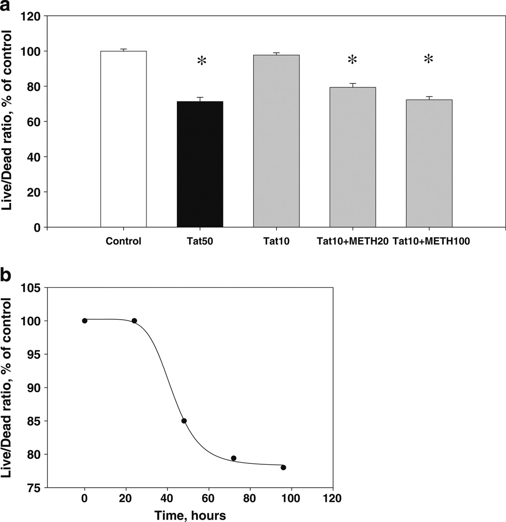
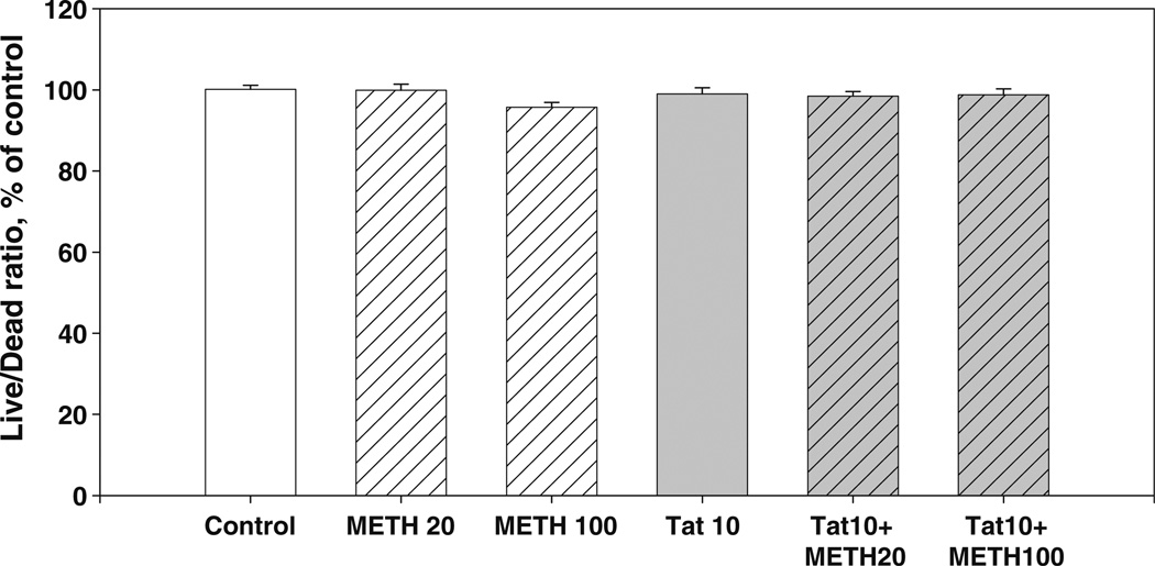
The interactive effects of HIV-1 infection and methamphetamine (METH) abuse in producing cognitive dysfunction represent a serious medical problem; however, the neural mechanisms underlying this interactive neurotoxicity remain elusive. In this study, we report that a combination of low, sub-toxic doses of METH + HIV-1 Tat 1-86 B, but not METH + HIV-1 gp120, directly induces death of rodent midbrain neurons in vitro. The effects of D1- and NMDA-receptor specific antagonists (SCH23390 and MK-801, respectively) on the neurotoxicity of different doses of METH or HIV-1 Tat alone and on the METH + HIV-1Tat interaction in midbrain neuronal cultures suggest that the induction of the cell death cascade by METH and Tat requires both dopaminergic (D1) and N-methyl D-aspartate (NMDA) receptor-mediated signaling. This interactive METH+Tat neurotoxicity does not occur in cultures of hippocampal neurons, which are predominately glutamatergic, express very low levels of dopamine receptors, and have no functional dopamine transporter (DAT). Thus, the presence of a subpopulation of neurons capable of dopamine release/uptake is essential for METH+Tat induction of the cell death cascade. Overall, our results support the hypothesis that METH and HIV-1 Tat disrupt the normal conjunction of signaling between D1 and NMDA receptors, resulting in neural dysfunction and death.


Conflict of interest statement

Conflict of interest The authors declare no conflict of interest.

Figures

Fig. 1
Fig. 1
Dose–response for METH toxicity in midbrain cell cultures. Cell viability measurements after 72-h treatment of midbrain cell cultures with varying concentrations (1 µM – 5 mM) of METH. Results (Live/Dead ratio, % of control) are presented as mean values ± SEM, n of sister cultures analyzed =8 per group. *- indicates the significant (P<0.05) differences in the Live/Dead ratios (compared to control cultures)
Fig. 2
Fig. 2
Combined sub-lethal doses of Tat + METH (a) or gp120+ METH (b) and cell viability. a Combined toxicity of 10 nM Tat+ 20 µM METH in midbrain cell cultures. Live/Dead ratios were measured after 72 h of treatment. Results (Live/Dead ratio, % of control) are presented as mean values ± SEM, n of sister cultures analyzed =8 per group. *- indicates the significant (P<0.05) differences in the Live/Dead ratios (compared to control cultures). b The effect of 30 pM gp120+20 µM METH after 72 h of treatment. Results (Live/Dead ratio, % of control) are presented as mean values ± SEM, n of sister cultures analyzed =8 per group. No significant changes in the Live/Dead ratios were observed
Fig. 3
Fig. 3
Cell viability decrements resulting from a toxic dose of Tat and a combination of non-toxic doses of Tat + METH. a The maximal cytotoxic effect (after 72 h of treatment) of 50 nM Tat and combinations of 10 nM dose of Tat with sub-lethal (20 and 100 µM) doses of METH in midbrain cell cultures. Results (Live/Dead ratio, % of control) are presented as mean values ± SEM, n of sister cultures analyzed =8 per group. *- indicates the significant (P<0.05) differences in the Live/Dead ratios (compared to control cultures). b The time course of Live/Dead ratio decline produced by 10 nM Tat+20 µM METH in midbrain cell cultures. Plot was best fit with a four-parameter sigmoid equation (R2>0.99)
Fig. 4
Fig. 4
Combined sub-lethal doses of Tat and METH and hippocampal cell viability. The effect of 10 nM Tat and (20 µM and 100 µM) METH after 72 h of treatment. Results (Live/Dead ratio, % of control) are presented as mean values ± SEM, n of sister cultures analyzed =8 per group. No significant changes in the Live/Dead ratios were observed in hippocampal cultures
Fig. 5
Fig. 5
Immunolocalization of NMDAR and D1R in midbrain neurons. Images of midbrain neurons showing colocalization of NR1 (green) and D1R (red), with Hoechst counterstained nuclei (blue). Midbrain cell cultures showed co-localization of NMDARs and D1Rs in cell bodies (left panels) and neuronal processes (right panels). Boxed selections were digitally magnified - scale bars: upper micrographs, 8 µm; lower micrographs, 20 µm
Fig. 6
Fig. 6
Inhibition of combined Tat + METH toxicity by the D1R-selective antagonist SCH23390. The protective effect of 10 µM SCH23390 against decreased midbrain cell viability after 72-h exposure to 10 nM Tat + 20 µM METH. Results (Live/Dead ratio, % of control) are presented as mean values ± SEM, n of sister cultures analyzed =8 per group. *- indicates the significant (P<0.05) differences in the Live/Dead ratios (compared to control cultures). SCH = SCH23390
Fig. 7
Fig. 7
Inhibition of combined Tat + METH toxicity by the NMDAR-specific antagonist MK-801. The protective effect of 0.1 and 1.0 µM MK-801 against decreased midbrain cell viability after 72-h exposure to 10 nM Tat+20 µM METH. Results (Live/Dead ratio, % of control) presented as mean values ± SEM, n of sister cultures analyzed =8 per group. *- indicates the significant (P<0.05) differences in the Live/Dead ratios (compared to control cultures). MK = MK-801

References

    1. Adams SM, Aksenova MV, Aksenov MY, Mactutus CF, Booze RM. ER-β mediates 17β-estradiol attenuation of HIV-1 Tat-induced apoptotic signaling. Synapse. 2010;64:829–838. - PMC - PubMed
    1. Aksenov MY, Aksenova MV, Nath A, Ray PD, Mactutus CF, Booze RM. Cocaine-mediated enhancement of Tat toxicity in rat hippocampal cell cultures: the role of oxidative stress and D1 dopamine receptor. Neurotoxicology. 2006;27(2):217–228. - PubMed
    1. Aksenov MY, Aksenova MV, Silvers JM, Mactutus CF, Booze RM. Different effects of selective dopamine uptake inhibitors, GBR 12909 and WIN 35428, on HIV-1 Tat toxicity in rat fetal midbrain neurons. Neurotoxicology. 2008;29(6):971–977. - PMC - PubMed
    1. Aksenov MY, Aksenova MV, Mactutus CF, Booze RM. Attenuated neurotoxicity of the transactivation-defective HIV-1 Tat protein in hippocampal cell cultures. Exp Neurol. 2009;219(2):586–590. - PMC - PubMed
    1. Aksenova MV, Silvers JM, Aksenov MY, Nath A, Ray PD, Mactutus CF, Booze RM. HIV-1 Tat neurotoxicity in primary cultures of rat midbrain fetal neurons: changes in dopamine transporter binding and immunoreactivity. Neurosci Lett. 2006;395(3):235–239. - PubMed

Publication types

MeSH terms

Substances