

Dual analysis for mycobacteria and propionibacteria in sarcoidosis BAL
- PMID: 22552860
- PMCID: PMC3526106
- DOI: 10.1007/s10875-012-9700-5
Dual analysis for mycobacteria and propionibacteria in sarcoidosis BAL
Abstract
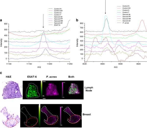
Purpose: Sarcoidosis is a non-caseating granulomatous disease for which a role for infectious antigens continues to strengthen. Recent studies have reported molecular evidence of mycobacteria or propionibacteria. We assessed for immune responses against mycobacterial and propionibacterial antigens in sarcoidosis bronchoalveolar lavage (BAL) using flow cytometry, and localized signals consistent with microbial antigens with sarcoidosis specimens, using matrix-assisted laser desorption ionization imaging mass spectrometry (MALDI-IMS).
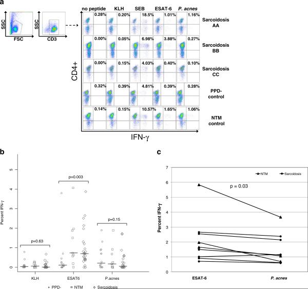
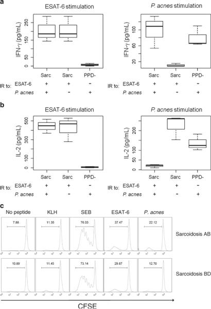
Methods: BAL cells from 27 sarcoidosis, 14 PPD- controls, and 9 subjects with nontuberculosis mycobacterial (NTM) infections were analyzed for production of IFN-γ after stimulation with mycobacterial ESAT-6 and Propionibacterium acnes proteins. To complement the immunological data, MALDI-IMS was performed to localize ESAT-6 and Propionibacterium acnes signals within sarcoidosis and control specimens.
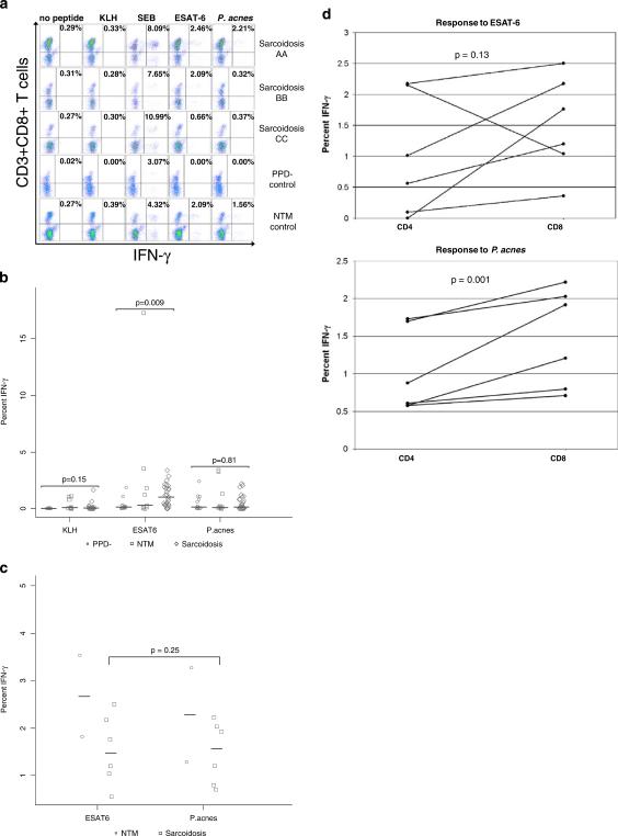
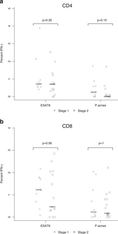
Results: CD4+ immunologic analysis for mycobacteria was positive in 17/27 sarcoidosis subjects, compared to 2/14 PPD- subjects, and 5/9 NTM subjects (p = 0.008 and p = 0.71 respectively, Fisher's exact test). There was no significant difference for recognition of P. acnes, which occurred only in sarcoidosis subjects that also recognized ESAT-6. Similar results were also observed for the CD8+ immunologic analysis. MALDI-IMS localized signals consistent with ESAT-6 only within sites of granulomatous inflammation, whereas P. acnes signals were distributed throughout the specimen.
Conclusions: MALDI-IMS localizes signals consistent with ESAT-6 to sarcoidosis granulomas, whereas no specific localization of P. acnes signals is detected. Immune responses against both mycobacterial and P. acnes are present within sarcoidosis BAL, but only mycobacterial signals are distinct from disease controls. These immunologic and molecular investigations support further investigation of the microbial community within sarcoidosis granulomas.
Figures
References
-
- Yanagisawa K, Shyr Y, Xu BJ, Massion PP, Larsen PH, White BC, et al. Proteomic patterns of tumour subsets in non-small-cell lung cancer. Lancet. 2003;362(9382):433–9. - PubMed
-
- Demirkok SS, Basaranoglu M, Coker E, Karayel T. Seasonality of the onset of symptoms, tuberculin test anergy and Kveim positive reaction in a large cohort of patients with sarcoidosis. Respirology. 2007;12(4):591–3. - PubMed
-
- Kajdasz DK, Judson MA, Mohr LC, Jr, Lackland DT. Geographic variation in sarcoidosis in South Carolina: its relation to socioeconomic status and health care indicators. Am J Epidemiol. 1999;150(3):271–8. - PubMed
Publication types
MeSH terms
Substances
Grants and funding
- 1 UL1 RR024975/RR/NCRR NIH HHS/United States
- R01 GM058008/GM/NIGMS NIH HHS/United States
- TL1 RR024978/RR/NCRR NIH HHS/United States
- 5R01-GM058008-11/GM/NIGMS NIH HHS/United States
- R01-AI65744/AI/NIAID NIH HHS/United States
- T32 HL069765/HL/NHLBI NIH HHS/United States
- P30 CA068485/CA/NCI NIH HHS/United States
- MO1 RR-00095/RR/NCRR NIH HHS/United States
- R01 AI065744/AI/NIAID NIH HHS/United States
- R01-HL83839/HL/NHLBI NIH HHS/United States
- KL2 RR024977/RR/NCRR NIH HHS/United States
- M01 RR000095/RR/NCRR NIH HHS/United States
- R01 HL083839/HL/NHLBI NIH HHS/United States
- 5P30-CA068485-13/CA/NCI NIH HHS/United States
- UL1 RR024975/RR/NCRR NIH HHS/United States
LinkOut - more resources
Full Text Sources
Other Literature Sources
Medical
Research Materials
