Review
. 2012 Nov;27(11):2039-2048.
doi: 10.1007/s00467-012-2175-z. Epub 2012 May 3.

Phosphate homeostasis and its role in bone health

Affiliations
Review

Phosphate homeostasis and its role in bone health

Maria Goretti Penido et al. Pediatr Nephrol. 2012 Nov.

Erratum in

Abstract

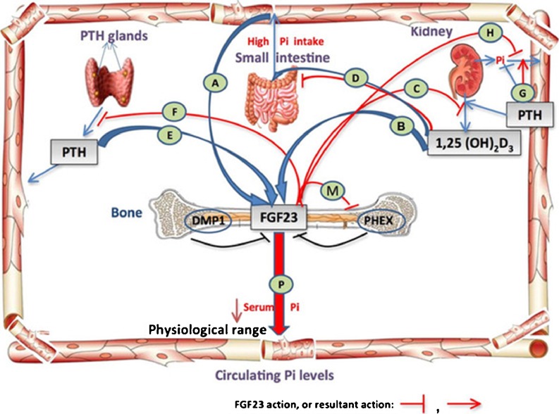
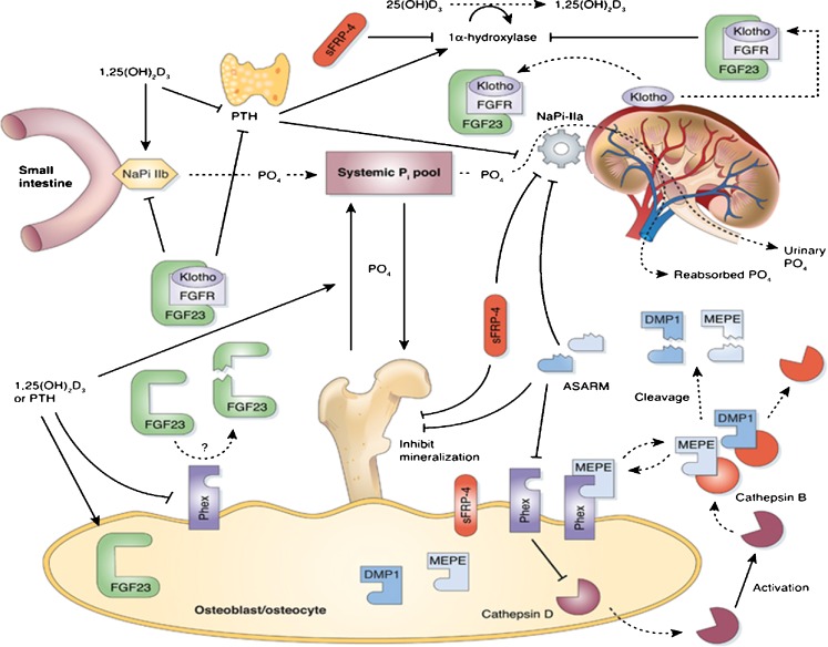
Phosphate is one of the most abundant minerals in the body, and its serum levels are regulated by a complex set of processes occurring in the intestine, skeleton, and kidneys. The currently known main regulators of phosphate homeostasis include parathyroid hormone (PTH), calcitriol, and a number of peptides collectively known as the "phosphatonins" of which fibroblast growth factor-23 (FGF-23) has been best defined. Maintenance of extracellular and intracellular phosphate levels within a narrow range is important for many biological processes, including energy metabolism, cell signaling, regulation of protein synthesis, skeletal development, and bone integrity. The presence of adequate amounts of phosphate is critical for the process of apoptosis of mature chondrocytes in the growth plate. Without the presence of this mineral in high enough quantities, chondrocytes will not go into apoptosis, and the normal physiological chain of events that includes invasion of blood vessels and the generation of new bone will be blocked, resulting in rickets and delayed growth. In the rest of the skeleton, hypophosphatemia will result in osteomalacia due to an insufficient formation of hydroxyapatite. This review will address phosphate metabolism and its role in bone health.


Figures

Fig. 1
Fig. 1
Regulatory mechanisms of phosphate homeostasis. A–H Intestine–bone–kidney–parathyroid axis. In the intestine, phosphate is actively absorbed through the cells or passively through the paracellular pathway. Its serum concentration is tightly regulated by parathyroid hormone (PTH), bone, and kidney. PTH stimulates phosphate excretion and calcitriol synthesis in the kidney; in turn, low phosphate and calcitriol directly inhibit PTH production. In the bone, PTH may stimulate fibroblast growth factor-23 (FGF-23) production and increase phosphate release following an increase in bone resorption. In the kidney, FGF-23 suppresses phosphate reabsorption and the synthesis of 1α, 25-dihydroxyvitamin D [1,25(OH)2D3], and in the parathyroid glands FGF23 suppresses the production and secretion of PTH. FGF23 is negatively regulated by bone signaling mechanisms (DMP1 and PHEX) and is positively regulated by systemic factors [serum inorganic phosphate (Pi), 1,25(OH)2D3, PTH (?)]. (Diagram is modified with permission from R. Sapir-Koren and G. Livshits: Bone mineralization and regulation of phosphate homeostasis. IBMS BoneKEy 8:286–300, 2011)
Fig. 2
Fig. 2
Phosphate transport in the intestine. The sodium-dependent phosphate transporters of the NaPi-IIb type are present at the luminal surface of the enterocyte (brush border membrane). NaPi-IIb transporters are electrogenic and have high affinity for inorganic phosphate (Pi). Energy for this transport process is provided by an inward downhill sodium gradient, maintained by transport of Na+ from the cell via a Na+/K+ ATPase cotransporter at the basolateral membrane. The phosphate incorporated into the enterocytes by this mechanism is transferred to the circulation by poorly understood mechanisms. Phosphate absorption also occurs via a sodium-independent process(es), such as diffusional movement across the intercellular spaces in the intestine. (Adapted from T.O. Carpentar: Chapter 10: Primary disorders of phosphate Mmtabolism, 2010, with permission from ENDOTEXT. Reprinted from www.endotext.org)
Fig. 3
Fig. 3
Phosphate transcellular transport in the proximal tubule. Phosphate movement from the renal tubule fluid to the peritubular capillary blood indicates that phosphate reabsorption is principally a unidirectional process that proceeds by a transcellular mechanism. Phosphate enters the tubular cell across the luminal membrane via a saturable, active, and sodium-dependent transport system. The rate of phosphate transport is dependent on the abundance of transporters functioning in the membrane and the magnitude of the Na+ gradient maintained across the luminal membrane, with the latter depending on the Na+/ATPase or sodium pump on the basolateral membrane. The rate-limiting step in phosphate transcellular transport is likely the Na+-dependent entry of inorganic phosphate (Pi) across the luminal membrane. Several phosphate transport systems have been postulated to explain the still poorly understood transport of phosphate across the basolateral membrane, including Na+–Pi cotransport via type III Na –Pi cotransporters, passive diffusion, and anion exchange. (Figure is adapted from Naderi and Reilly [11] and reprinted with permission from Macmillan Publishers)
Fig. 4
Fig. 4
Overall picture of the physiology of FGF-23 and its role in phosphate homeostasis. FGF-23 inhibits renal Pi reabsorption by reducing the apical expression and activity of NaPi-IIa in the proximal tubule epithelium. FGF-23 also reduces intestinal absorption of dietary Pi through a vitamin D receptor (VDR)-dependent decrease in NaPi-IIb activity. The latter phenomenon is most likely secondary to FGF-23-mediated reduction of circulating 1,25(OH)2D3 synthesis through suppression of 1α(OH)ase expression in the kidney. The proteins required for mineralization [osteocalcin, bone sialoprotein, vitronectin, and dentin matrix protein 1 (DMP-1)] are in dynamic balance with the ASARM peptide, an inhibitor of mineralization. In vitro, data indicates that PHEX-sequestered matrix extracellular phosphoglycoprotein (MEPE) is protected from matrix proteases. The PHEX sequestration is likely reversible, and free unbound-MEPE is vulnerable to cleavage by extracellular matrix proteases/cathepsin B. Cleavage of MEPE would result in the release of the protease-resistant stable ASARM peptide (aspartic acid-rich motif). Free ASARM peptide may also sterically inhibit the NPT2 co-transporter by direct binding. PHEX is expressed in the parathyroid glands and may have a role in the normal regulation of PTH. The full-length uncleaved FGF-23 molecule may indirectly/directly downregulate the proteins required for mineralization. Thus, FGF-23 and/or other cytokines are likely directly/indirectly responsible for the changes in expression of these proteins. FGF-23 indirectly or directly suppresses expression of the NPT2 phosphate co-transporter, suppresses expression of 1α-hydroxylase, and increases expression of 24-hydroxylase [decrease in 1,25(OH)2D3]. FGF-23 and Klotho might function in a common single transduction pathway. (Modified from R. Pawel et al.: Recent advances in the renal–skeletal–gut axis that controls phosphate homeostasis. Lab Invest 89:7–14, 2009. Reprinted by permission from Macmillian Publishers)

Similar articles

Cited by

References

    1. Marks J, Edward S, Debnam ES, Unwi RJ. Phosphate homeostasis and the renal-gastrointestinal axis. Am J Physiol Renal Physiol. 2010;299:F285–F296. doi: 10.1152/ajprenal.00508.2009. - DOI - PubMed
    1. Berndt T, Thomas LF, Craig TA, Sommer S, Li X, Bergstralh EJ, Kumar R. Evidence for a signaling axis by which intestinal phosphate rapidly modulates renal phosphate reabsorption. Proc Natl Acad Sci USA. 2007;104:11085–11090. doi: 10.1073/pnas.0704446104. - DOI - PMC - PubMed
    1. Shaikh A, Berndt T, Kumar R. Regulation of phosphate homeostasis by the phosphatonins and other novel mediators. Pediatr Nephrol. 2008;23:1203–1210. doi: 10.1007/s00467-008-0751-z. - DOI - PMC - PubMed
    1. Giral H, Caldas Y, Sutherland E, Wilson P, Breusegem SY, Barry N, Blaine J, Jiang T, Wang XX, Levi M. Regulation of the rat intestinal Na-dependent phosphate transporters by dietary phosphate. Am J Physiol Renal Physiol. 2009;297:F1466–F1475. doi: 10.1152/ajprenal.00279.2009. - DOI - PMC - PubMed
    1. Reining SC, Liesegang A, Betz H, Biber J, Murer H, Hernando N. Expression of renal and intestinal Na/Pi cotransporters in the absence of GABARAP. Pflügers Arch. 2010;460:201–217. doi: 10.1007/s00424-010-0832-2. - DOI - PubMed

MeSH terms

LinkOut - more resources