

Human cytomegalovirus UL97 kinase alters the accumulation of CDK1
- PMID: 22552942
- PMCID: PMC3541764
- DOI: 10.1099/vir.0.039214-0
Human cytomegalovirus UL97 kinase alters the accumulation of CDK1
Abstract
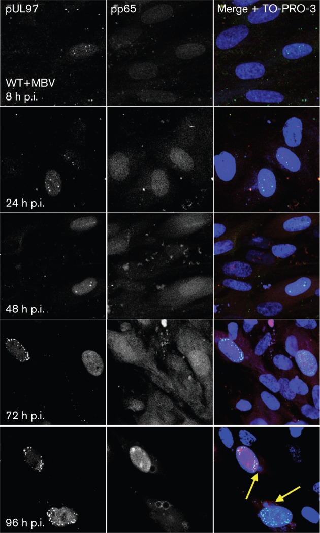
The UL97 protein kinase is a serine/threonine kinase expressed by human cytomegalovirus (CMV) that phosphorylates ganciclovir. An investigation of the subcellular localization of pUL97 in infected cells indicated that, early in infection, pUL97 localized to focal sites in the nucleus that transitioned to subnuclear compartments and eventually throughout the entire nucleus. When UL97 kinase activity was eliminated with a K355M mutation or pharmacologically inhibited with maribavir, the expansion and redistribution of pUL97 foci within the nucleus was delayed, nuclear reorganization did not occur and assembly complexes in the cytoplasm failed to form normally. As UL97 kinase and its homologues appear to be functionally related to CDK1, a known regulator of nuclear structural organization, the effects of the UL97 kinase on CDK1 were investigated. Expression of CDK1 in infected cells appeared to be induced by UL97 kinase activity at the level of transcription and was not tied to other virus life-cycle events, such as viral DNA replication or virion assembly. These results suggest that, in addition to phosphorylating CDK1 targets, the UL97 kinase modifies G₂/M cell-cycle checkpoint regulators, specifically CDK1, to promote virus replication.
Figures


Similar articles
-
Comparison between human cytomegalovirus pUL97 and murine cytomegalovirus (MCMV) pM97 expressed by MCMV and vaccinia virus: pM97 does not confer ganciclovir sensitivity.J Virol. 2000 Nov;74(22):10729-36. doi: 10.1128/jvi.74.22.10729-10736.2000. J Virol. 2000. PMID: 11044117 Free PMC article.
-
The human cytomegalovirus UL97 protein is phosphorylated and a component of virions.Virology. 1997 Apr 28;231(1):72-80. doi: 10.1006/viro.1997.8523. Virology. 1997. PMID: 9143304
-
Viral mimicry of Cdc2/cyclin-dependent kinase 1 mediates disruption of nuclear lamina during human cytomegalovirus nuclear egress.PLoS Pathog. 2009 Jan;5(1):e1000275. doi: 10.1371/journal.ppat.1000275. Epub 2009 Jan 23. PLoS Pathog. 2009. PMID: 19165338 Free PMC article.
-
Function of human cytomegalovirus UL97 kinase in viral infection and its inhibition by maribavir.Rev Med Virol. 2009 Jul;19(4):215-29. doi: 10.1002/rmv.615. Rev Med Virol. 2009. PMID: 19434630 Free PMC article. Review.
-
Cytomegalovirus UL97 mutations in the era of ganciclovir and maribavir.Rev Med Virol. 2008 Jul-Aug;18(4):233-46. doi: 10.1002/rmv.574. Rev Med Virol. 2008. PMID: 18383425 Review.
Cited by
-
How SARS-CoV-2 and Other Viruses Build an Invasion Route to Hijack the Host Nucleocytoplasmic Trafficking System.Cells. 2021 Jun 7;10(6):1424. doi: 10.3390/cells10061424. Cells. 2021. PMID: 34200500 Free PMC article. Review.
-
Strategies for the Viral Exploitation of Nuclear Pore Transport Pathways.Viruses. 2025 Jan 23;17(2):151. doi: 10.3390/v17020151. Viruses. 2025. PMID: 40006906 Free PMC article. Review.
-
Conserved Herpesvirus Protein Kinases Target SAMHD1 to Facilitate Virus Replication.Cell Rep. 2019 Jul 9;28(2):449-459.e5. doi: 10.1016/j.celrep.2019.04.020. Cell Rep. 2019. PMID: 31291580 Free PMC article.
-
Differential properties of cytomegalovirus pUL97 kinase isoforms affect viral replication and maribavir susceptibility.J Virol. 2014 May;88(9):4776-85. doi: 10.1128/JVI.00192-14. Epub 2014 Feb 12. J Virol. 2014. PMID: 24522923 Free PMC article.
-
Proteomic analysis of the multimeric nuclear egress complex of human cytomegalovirus.Mol Cell Proteomics. 2014 Aug;13(8):2132-46. doi: 10.1074/mcp.M113.035782. Epub 2014 Jun 26. Mol Cell Proteomics. 2014. PMID: 24969177 Free PMC article.
References
-
- Anon. (2007). Maribavir: 1263W94, Benzimidavir, GW 1263, GW 1263W94, VP41263. Drugs R D 8, 188–192 - PubMed
-
- Advani S. J., Brandimarti R., Weichselbaum R. R., Roizman B. (2000). The disappearance of cyclins A and B and the increase in activity of the G2/M-phase cellular kinase cdc2 in herpes simplex virus 1-infected cells require expression of the α22/US1.5 and UL13 viral genes. J Virol 74, 8–15 10.1128/JVI.74.1.8-15.2000 - DOI - PMC - PubMed
-
- Arcangeletti M. C., De Conto F., Ferraglia F., Pinardi F., Gatti R., Orlandini G., Calderaro A., Motta F., Medici M. C. & other authors (2003). Human cytomegalovirus proteins PP65 and IEP72 are targeted to distinct compartments in nuclei and nuclear matrices of infected human embryo fibroblasts. J Cell Biochem 90, 1056–1067 10.1002/jcb.10655 - DOI - PubMed
Publication types
MeSH terms
Substances
Grants and funding
LinkOut - more resources
Full Text Sources
Research Materials
Miscellaneous