Review
. 2013 Jan;228(1):9-20.
doi: 10.1002/jcp.24104.

The abbreviated pluripotent cell cycle

Affiliations
Review

The abbreviated pluripotent cell cycle

Kristina Kapinas et al. J Cell Physiol. 2013 Jan.

Abstract

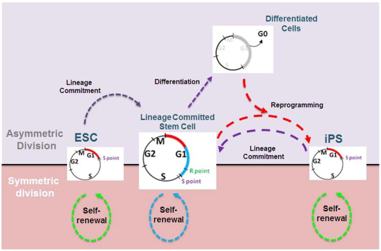
Human embryonic stem cells (hESCs) and induced pluripotent stem cells proliferate rapidly and divide symmetrically producing equivalent progeny cells. In contrast, lineage committed cells acquire an extended symmetrical cell cycle. Self-renewal of tissue-specific stem cells is sustained by asymmetric cell division where one progeny cell remains a progenitor while the partner progeny cell exits the cell cycle and differentiates. There are three principal contexts for considering the operation and regulation of the pluripotent cell cycle: temporal, regulatory, and structural. The primary temporal context that the pluripotent self-renewal cell cycle of hESCs is a short G1 period without reducing periods of time allocated to S phase, G2, and mitosis. The rules that govern proliferation in hESCs remain to be comprehensively established. However, several lines of evidence suggest a key role for the naïve transcriptome of hESCs, which is competent to stringently regulate the embryonic stem cell (ESC) cell cycle. This supports the requirements of pluripotent cells to self-propagate while suppressing expression of genes that confer lineage commitment and/or tissue specificity. However, for the first time, we consider unique dimensions to the architectural organization and assembly of regulatory machinery for gene expression in nuclear microenviornments that define parameters of pluripotency. From both fundamental biological and clinical perspectives, understanding control of the abbreviated ESC cycle can provide options to coordinate control of proliferation versus differentiation. Wound healing, tissue engineering, and cell-based therapy to mitigate developmental aberrations illustrate applications that benefit from knowledge of the biology of the pluripotent cell cycle.


Conflict of interest statement

The authors have no conflicts of interest.

Figures

Figure 1
Figure 1. Differences between pluripotent and lineage committed cell cycle length
The four stages of the somatic cell cycle (G1: gap 1, S: synthesis of DNA, G2: gap 2, and M: mitosis) support duplication of the genome and subsequent segregation of a diploid set of chromosomes into two progeny cells. Post-fertilization, during early development, embryonic stem cells (ESC), derived from the inner cell mass (ICM) of blastocysts, have an abbreviated cell cycle, as a consequence of a very short G1 phase (red continuous segment; 2-3 h). During lineage commitment, ESCs undergo asymmetric division to produce more stem cells and new precursor cells (lineage committed stem cells and lineage specific progenitors) that will support growth, differentiation, and organogenesis. These precursor cells continue dividing with a normal cell cycle, but extended G1 phase (red/blue continuous segments; 8-12 h). The reprogramming of pre-committed somatic cells to a pluripotent state results in the reacquisition of a shortened cell cycle with a short G1 phase (red continuous segment; 2-3 h) and constant S, G2 and M phases. Upon differentiation, the cell exits the cell cycle after mitosis and goes to G0 (gray continuous segment). All pluripotent cells have the potential to undergo symmetric or asymmetric division. This equilibrated state (central black line) allows the cells to be responsive to signals for self-renewal or lineage commitment. The green and blue dotted lines depict cell divisions during self-renewal. The purple dotted lines represent the intermediate steps necessary for lineage commitment. The red dotted lines symbolize the chain of events necessary to reprogram cells (iPS cells). For simplicity, the intermediary states between lineage-committed stem cells and differentiated cells are not shown. R and S points represent the restriction points controlling the G1/S transition. Note, that the R point is only present in committed cells. Both restriction points are more extensively described in Figure 5.
Figure 2
Figure 2. Multiple checkpoints control cell cycle progression
(a) The cell cycle is regulated by several critical cell cycle checkpoints (ticks) at which competency for cell cycle progression is monitored. Entry into and exit from the cell cycle (black lines and lettering) is controlled by growth regulatory factors (e.g. cytokines, growth factors, cell adhesion and/or cell–cell contact), which determine self-renewal of stem cells and expansion of pre-committed progenitor cells. The biochemical parameters associated with each cell cycle checkpoint are indicated by red lettering. Options for defaulting to apoptosis (yellow lettering) during G1 and G2 are evaluated by surveillance mechanisms that assess fidelity of structural and regulatory parameters of cell cycle control. (b) Transcription factors (green) are organized in distinct foci in the interphasic nuclei. Although some lineage-specific transcription factors (i.e. RUNX2) are retained on target gene promoters in chromosomes at all stages of mitosis, others do not associate with chromosomes and are degraded. This retention of transcription factors in addition to the occurrence of certain histone modifications indicate that certain genes are bookmarked for expression after mitosis.
Figure 3
Figure 3. Surveillance and editing mechanisms mediating checkpoint control
(a) Surveillance mechanisms monitor multiple biochemical and architectural parameters that control cell cycle progression. These parameters include the intracellular levels of regulatory proteins, structural and informational integrity of the genome, as well as extracellular signals governing cell cycle progression. The integration of this regulatory input can result in (i) competency for cell cycle progression (green traffic light and arrows), (ii) cell cycle inhibition and activation of editing mechanisms (yellow traffic light and arrows) or (iii) the active and regulated destruction of the cell in response to apoptotic signals (red traffic light and red arrow). (b) Traverse of the cell cycle is regulated by a series of checkpoints at strategic positions within the cell cycle. Several major checkpoints (yellow arrows with ticks and light purple lettering) only allow a cell to commit to a subsequent cell cycle stage upon satisfying essential biochemical and architectural criteria governing competency for cell cycle progression (green traffic lights). For example, at the ‘restriction point’ surveillance mechanisms (yellow traffic lights) integrate cell growth stimulatory and inhibitory signals, including growth factors, cell adhesion and nutrient status (light purple lettering). Checkpoints in G1 and G2 are necessary to ensure the integrity of the genome and, if necessary, activate chromatin editing mechanisms (light purple lettering). The spindle assembly checkpoint ensures equal chromosome segregation. (c) Checkpoint control mechanisms monitor intracellular levels of cell cycle regulatory factors, as well as parameters of chromatin architecture. For example, the activation of cyclin-dependent kinases reflects the sensing of intracellular concentrations of the cognate cyclins. CDK activation is attenuated by CDK inhibitor proteins (CKIs) which inactivate CDK/cyclin complexes. Competency for cell cycle progression requires that cyclin levels reach a threshold (e.g. by exceeding the levels of available CKIs, or phosphorylation events altering the affinities of cyclins and CKIs for CDKs). As a consequence, activated CDK/cyclin complexes phosphorylate transcription factors that regulate expression of cell cycle stage-specific genes. Furthermore, key checkpoints in G1 and G2 monitor chromatin integrity and perform essential editing functions. DNA damage activates DNA-repair mechanisms that fix informational errors in the genome and restore nucleosomal organization by chromatin remodeling.
Figure 4
Figure 4. (A) Multiple Levels of Nuclear Organization
The organization of cognate DNA-regulatory elements in a linear fashion within gene promoters comprises the primary level of nuclear organization. The distance between these regulatory sites is additionally regulated by the folding of DNA into nucleosomes and higher order chromatin structures. Scaffolding nuclear proteins, usually transcription factors (TF), assemble multiprotein complexes to facilitate the combinatorial control of gene expression within the context of nuclear structure, thus forming dynamic microenvironments within the nucleus. Typical nuclear microenvironments contain various co-regulatory proteins that are involved in combinatorial control of gene activation, as well as repression, chromatin remodeling and cellular signaling. In addition, many nuclear microenvironments are equally partitioned during mitosis to epigenetically regulate cell growth and phenotypic properties.
Figure 5
Figure 5. Transcriptional control at the G1/S phase transition
The genes encoding cell cycle regulatory subunits (e.g. cyclin E) and histone biosynthesis (e.g. H4) are each controlled by intricate arrays of promoter regulatory elements that influence transcriptional initiation by RNA polymerase II. E2F elements in the promoter of the cyclin E gene interact with E2F factors that associate with CDKs, cyclins and pRB-related proteins (R-point). In contrast, histone genes are controlled by the site II cell cycle regulatory element, which interacts with CDP-cut and IRF2 proteins, and the HINFP/p220NPAT complex. Analogous to E2Fdependent mechanisms, CDP-cut interacts with CDK1, cyclin A and pRB, whereas IRF2 performs an activating function similar to ‘free’ E2F. HINFP binds to this cell cycle regulatory element (site II) and recruits p220NPAT, thus integrating signals from the cyclin E/CDK2 kinase pathway (S-point). The presence of SP1 in the promoters of G1/S phase-related genes provides a shared mechanism for further enhancement of transcription at the onset of S phase.
Figure 6
Figure 6. The cell cycle control of histone gene expression is required to support the abbreviated cell cycle in pluripotent stem cells
In pluripotent stem cells, as in lineage committed cells, histone genes are not regulated by an E2F/RB switch but by a HiNF-P/p220NPAT co-activation complex. However, the cell cycle dependent organization of p220NPAT foci is different between somatic and pluripotent stem cells. For example, in pluripotent stem cells, the number of p220NPAT foci increases in G1 prior to the CDK dependent phosphorylation of p220NPAT in the S phase (a, b; top panel). In contrast, the number of p220NPAT foci double from two to four upon entry into S phase in lineage committed cells (a, b; bottom panel). The increase in the kinetic of HLBs formation in pluripotent stem cells may render the p220NPAT/HiNF-P/histone gene regulatory complex poised for rapid activation by cyclin/CDK complexes to induce histone gene expression at the onset of DNA synthesis. (a) Diagrammatic representation of temporally different assembly of Histone Locus Bodies (HLBs) during G1/S transition in pluripotent vs lineage committed cells. (b) Immunofluorescence microscopy demonstrating temporal differences in the assembly of HLBs during G1/S transition in pluripotent vs lineage committed cells. Mitotically synchronized hES cells at various cell cycle stages were monitored by immunofluorescence (IF) microscopies for association of NPAT (green), showing spatial and temporal linkage of the histone gene cluster at 6p22 (red). DAPI staining (blue) was used to visualize the nucleus.

Similar articles

Cited by

References

    1. Adamo A, Sese B, Boue S, Castano J, Paramonov I, Barrero MJ, Izpisua Belmonte JC. LSD1 regulates the balance between self-renewal and differentiation in human embryonic stem cells. Nat Cell Biol. 2011;13:652–659. - PubMed
    1. Ahmed K, Dehghani H, Rugg-Gunn P, Fussner E, Rossant J, Bazett-Jones DP. Global chromatin architecture reflects pluripotency and lineage commitment in the early mouse embryo. PLoS ONE. 2010;5:e10531. - PMC - PubMed
    1. Amente S, Bertoni A, Morano A, Lania L, Avvedimento EV, Majello B. LSD1-mediated demethylation of histone H3 lysine 4 triggers Myc-induced transcription. Oncogene. 2010a;29:3691–3702. - PubMed
    1. Amente S, Lania L, Avvedimento EV, Majello B. DNA oxidation drives Myc mediated transcription. Cell Cycle. 2010b;9:3002–3004. - PubMed
    1. Amit M, Carpenter MK, Inokuma MS, Chiu CP, Harris CP, Waknitz MA, Itskovitz-Eldor J, Thomson JA. Clonally derived human embryonic stem cell lines maintain pluripotency and proliferative potential for prolonged periods of culture. Dev Biol. 2000;227:271–278. - PubMed

Publication types

LinkOut - more resources