Comparative Study
. 2012 May;29(5):449-59.
doi: 10.1002/da.21892.

Brain activation to facial expressions in youth with PTSD symptoms

Affiliations
Comparative Study

Brain activation to facial expressions in youth with PTSD symptoms

Amy S Garrett et al. Depress Anxiety. 2012 May.

Abstract

Objective: This study examined activation to facial expressions in youth with a history of interpersonal trauma and current posttraumatic stress symptoms (PTSS) compared to healthy controls (HC).

Design and analysis: Twenty-three medication-naive youth with PTSS and 23 age- and gender-matched HC underwent functional magnetic resonance imaging (fMRI) while viewing fearful, angry, sad, happy, and neutral faces. Data were analyzed for group differences in location of activation, as well as timing of activation during the early versus late phase of the block. Using SPM5, significant activation (P < .05 FWE [Family-Wise Error] corrected, extent = 10 voxels) associated with the main effect of group was identified. Activation from selected clusters was extracted to SPSS software for further analysis of specific facial expressions and temporal patterns of activation.

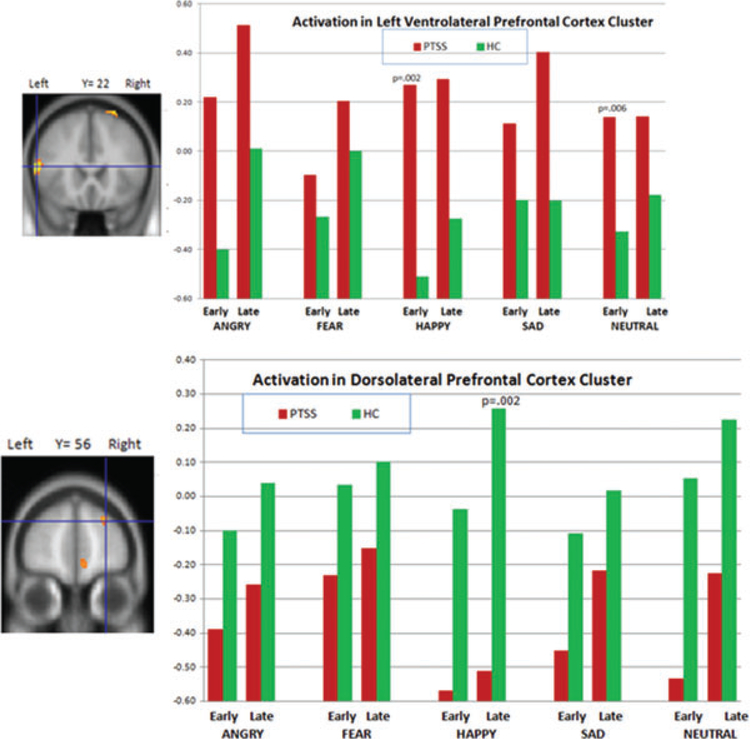
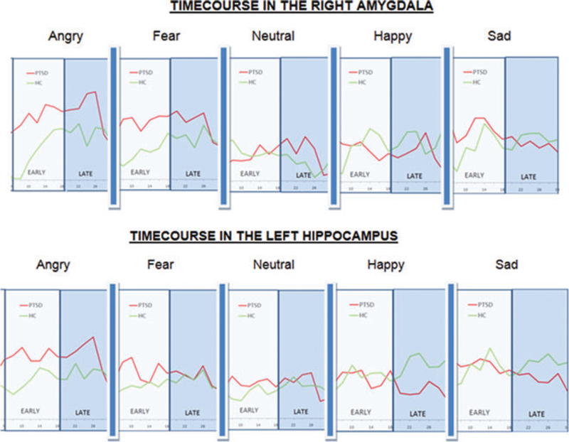
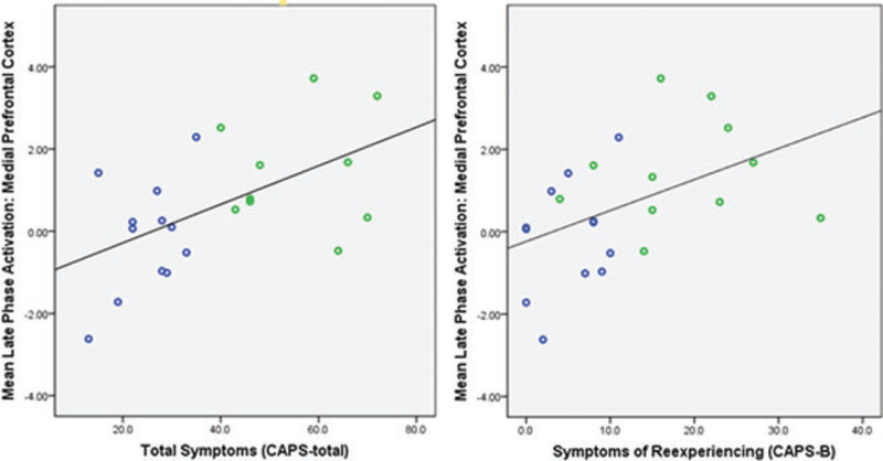
Results: The PTSS group showed significantly greater activation than controls in several regions, including the amygdala/hippocampus, medial prefrontal cortex, insula, and ventrolateral prefrontal cortex, and less activation than controls in the dorsolateral prefrontal cortex (DLPFC). These group differences in activation were greatest during angry, happy, and neutral faces, and predominantly during the early phase of the block. Post hoc analyses showed significant Group × Phase interactions in the right amygdala and left hippocampus.

Conclusions: Traumatic stress may impact development of brain regions important for emotion processing. Timing of activation may be altered in youth with PTSS.


Conflict of interest statement

Conflict of interest: None declared.

Figures

Figure 1.
Figure 1.
Main effect of group broken down by facial expression and phase. Significant group differences in left amygdala/hippocampus activation (at crosshairs) were greatest in the late phase of angry faces, and early and late phases of neutral faces; group differences in activation of the medial prefrontal cluster (at crosshairs) were greatest in the early phase of angry faces; group differences in activation in the insula were greatest during the early phase of angry faces, and early phase of neutral faces; group differences in activation in the left ventrolateral cluster (at crosshairs) were greatest during the early phase of happy faces, and the early phase of neutral faces; group differences in activation in the dorsolateral prefrontal cortex (DLPFC) were greatest in the late phase of happy faces.
Figure 1.
Figure 1.
Main effect of group broken down by facial expression and phase. Significant group differences in left amygdala/hippocampus activation (at crosshairs) were greatest in the late phase of angry faces, and early and late phases of neutral faces; group differences in activation of the medial prefrontal cluster (at crosshairs) were greatest in the early phase of angry faces; group differences in activation in the insula were greatest during the early phase of angry faces, and early phase of neutral faces; group differences in activation in the left ventrolateral cluster (at crosshairs) were greatest during the early phase of happy faces, and the early phase of neutral faces; group differences in activation in the dorsolateral prefrontal cortex (DLPFC) were greatest in the late phase of happy faces.
Figure 2.
Figure 2.
Time-courses of activation in the right amygdala (top) and left hippocampus (bottom) displaying a significant Group × Phase interaction. The posttraumatic stress symptoms (PTSS) group time-course is shown in red and the HC group is shown in green. The x-axis is seconds, and the early phase (0–12 s) and late phase (12–24 s) are indicated (both shifted 6 s forward to account for the hemodynamic lag). For the right amygdala, the PTSS activation is greater than the HC particularly in the early phases of the angry and fear blocks. For the left hippocampus, PTSS activation is greater during early and late phase of the angry blocks, but less during the late phase of the happy and sad blocks.
Figure 3.
Figure 3.
Significant correlation between CAPS score and activation in the ventral medial prefrontal cortex during the late phase of the fearful faces block: for total CAPS score, ρ = .55, P = .008; for the CAPS-B (re-experiencing ) subscale (ρ = .54, P = .008). Green datapoints indicate subjects with a full PTSD diagnosis, whereas blue are subthreshold.

References

    1. Copeland WE, et al. Traumatic events and posttraumatic stress in childhood. Arch Gen Psychiatry 2007;64(5):577–584. - PubMed
    1. Carrion VG, et al. Diurnal salivary cortisol in pediatric posttraumatic stress disorder. Biol Psychiatry 2002;51(7):575–582. - PubMed
    1. Carrion VG, et al. Toward an empirical definition of pediatric PTSD: the phenomenology of PTSD symptoms in youth. J Am Acad ChildAdolesc Psychiatry 2002;41(2):166–173. - PubMed
    1. King NJ, et al. Treating sexually abused children with posttraumatic stress symptoms: a randomized clinical trial. J Am Acad ChildAdolesc Psychiatry 2000;39(11):1347–1355. - PubMed
    1. Jackowski AP, et al. Neurostructural imaging findings in children with post-traumatic stress disorder: brief review. Psychiatry Clin Neurosci 2009;63(1):1–8. - PMC - PubMed

Publication types