. 2012 May 2;32(18):6231-9.
doi: 10.1523/JNEUROSCI.2543-11.2012.

Stimulus site and modality dependence of functional activity within the human spinal cord

Affiliations

Stimulus site and modality dependence of functional activity within the human spinal cord

Jonathan C W Brooks et al. J Neurosci. .

Abstract

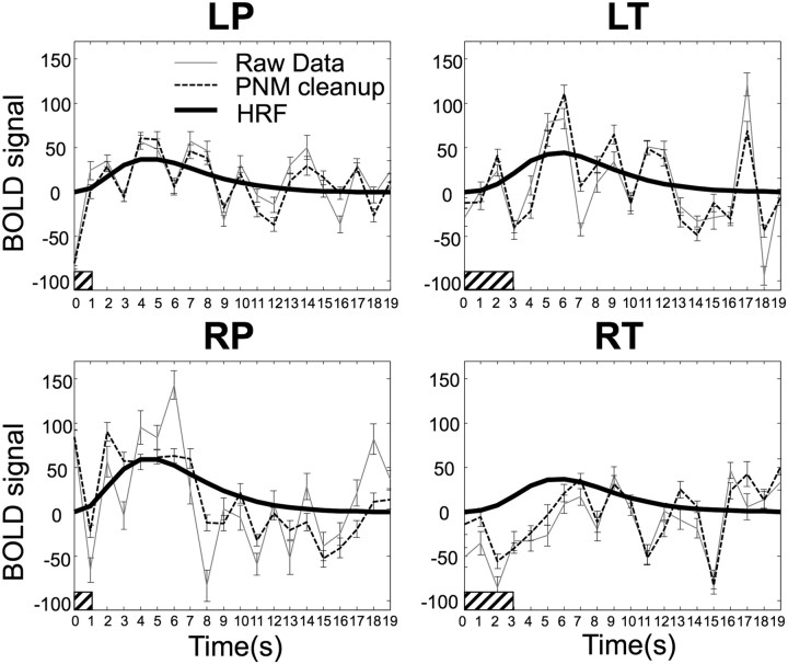
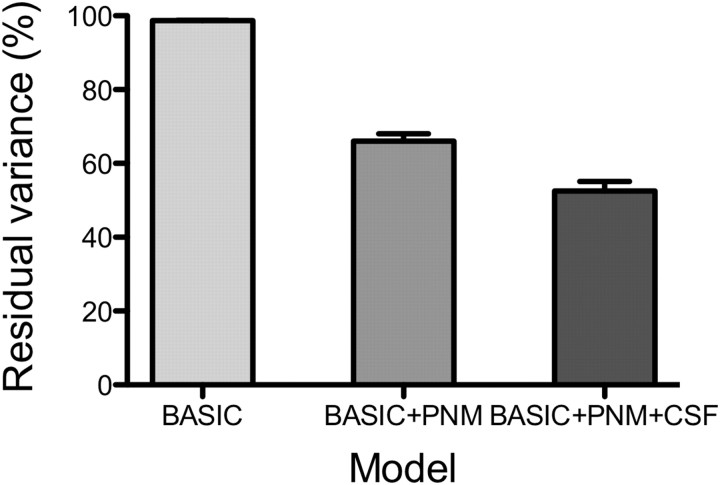
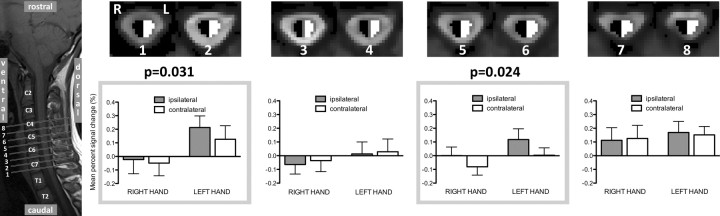
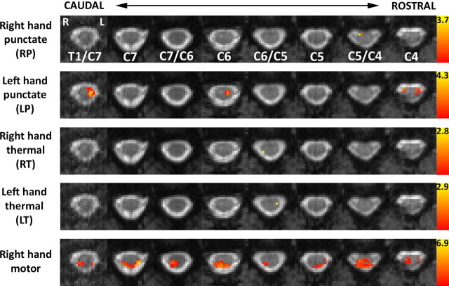
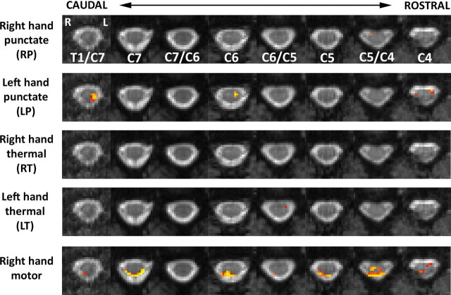
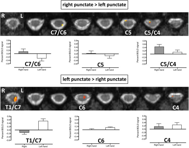
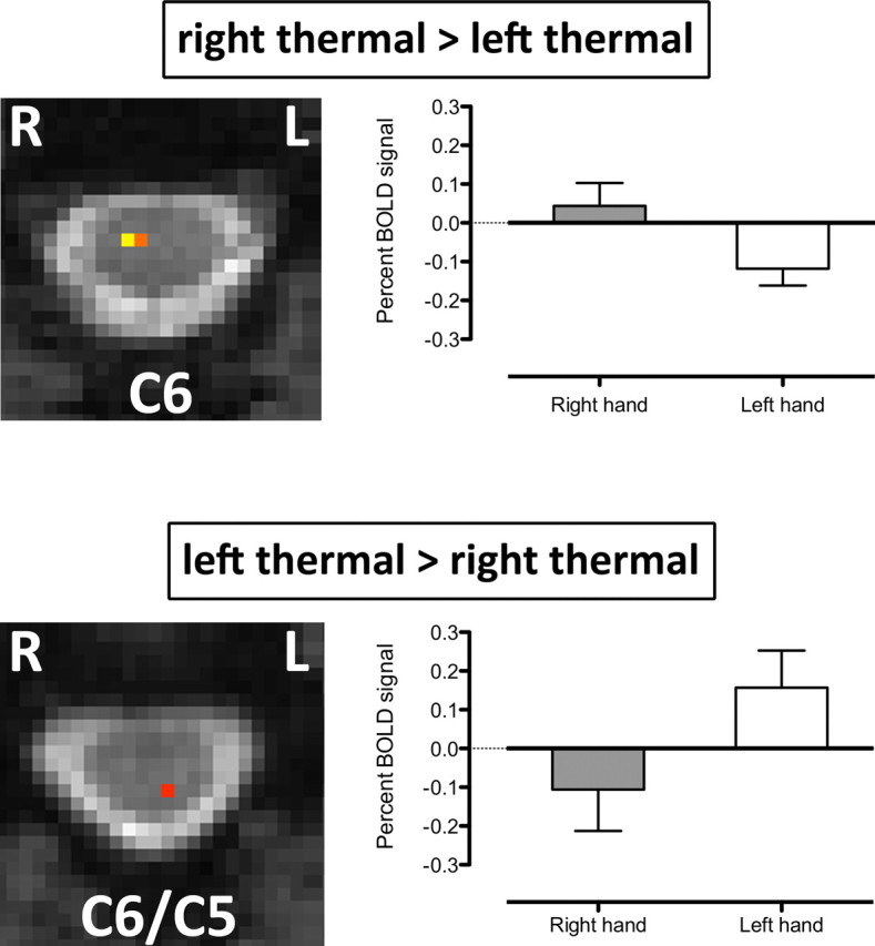
Chronic pain is thought to arise because of maladaptive changes occurring within the peripheral nervous system and CNS. The transition from acute to chronic pain is known to involve the spinal cord (Woolf and Salter, 2000). Therefore, to investigate altered human spinal cord function and translate results obtained from other species, a noninvasive neuroimaging technique is desirable. We have investigated the functional response in the cervical spinal cord of 18 healthy human subjects (aged 22-40 years) to noxious thermal and non-noxious tactile stimulation of the left and right forearms. Physiological noise, which is a significant source of signal variability in the spinal cord, was accounted for in the general linear model. Group analysis, performed using a mixed-effects model, revealed distinct regions of activity that were dependent on both the side and the type of stimulation. In particular, thermal stimulation on the medial aspect of the wrist produced activity within the C6/C5 segment ipsilateral to the side of stimulation. Similar to data recorded in animals (Fitzgerald, 1982), painful thermal stimuli produced increased ipsilateral and decreased contralateral blood flow, which may reflect, respectively, excitatory and inhibitory processes. Nonpainful punctate stimulation of the thenar eminence provoked more diffuse activity but was still ipsilateral to the side of stimulation. These results present the first noninvasive evidence for a lateralized response to noxious and non-noxious stimuli in the human spinal cord. The development of these techniques opens the path to understanding, at a subject-specific level, central sensitization processes that contribute to chronic pain states.


Figures

Figure 1.
Figure 1.
Group average (N = 18) peristimulus plots for each of the four applied stimuli (RP, LP, RT, and LT) taken from the peak voxel for each contrast in the group analysis. For each of the four peristimulus plots, the raw data are shown by the light solid line, and the time course after “PNM cleanup” is shown by the light dashed line. Error bars represent SEs. In general, the data after cleanup have fewer large jumps and look smoother, reflecting the reduction in data variation. The heavy solid line is the fitted hemodynamic response curve to the data after cleanup, the filled bar at the origin indicates the timing and duration of the applied stimulus, and HRF = haemodynamic response function.
Figure 2.
Figure 2.
Average variance reduction for three applied models (basic, basic + PNM, and basic + PNM + CSF) measured from voxels in the spinal cord. The percentage reduction in variance is relative to the baseline variance of the raw data and is expressed as a percentage of the nominal value (100%). Applying the basic design (experimental design only) reduced the variance by <2%, applying a PNM-reduced variance by a further 33%, and including a CSF regressor in addition to the PNM reduced the overall variance by 47.5%. The basic + PNM + CSF model was used to estimate activation in this study.
Figure 3.
Figure 3.
ROI analysis of spinal cord activation in response to punctate stimulation. BOLD percentage signal change was extracted from hand-drawn ROIs covering the dorsal and ventral regions of the right (black) and left (white) hemicords. The location of the relevant slices is shown on a sagittal section spanning from T1/C7 to C4 (i.e., covering the dorsal roots corresponding to dermatomes stimulated in this experiment). Averaged signal from adjacent slices was plotted (bar plots) for each stimulus location and each ROI, where bar shading corresponds to ipsilateral (filled bars) and contralateral (open bars) and thus refers to different sides of the cord, depending on the site of stimulation. A main effect of laterality was found for slices 1 and 2 and for slices 5 and 6 (repeated-measures ANOVA, p < 0.05).
Figure 4.
Figure 4.
Mixed-effects group results for punctate/thermal stimuli and rating task. Activation data, thresholded at “corrected” p < 0.003 (corresponding to Z > 2.75), are superimposed on the corresponding slices of the average EPI data from the standard cord. Activation was found for all contrasts and tended to be located ipsilaterally to the side of thermal or punctate stimulation. Activity during the four separate rating periods was averaged and constituted a right-hand sensory/motor task. Activity was located primarily within the ipsilateral hemicord and extended from dorsal (sensory) to ventral (motor) areas. Note that activity on the CSF/cord boundary was found exclusively on the dorsal cord surface and tended to be bilateral. Data are shown in radiological format (left side of cord on right side of each slice), and the level of each slice is shown relative to the vertebral body to which it is opposite (or at the midpoint between two vertebrae if that is where it lies).
Figure 5.
Figure 5.
Group results for punctate/thermal stimuli and rating task, subjected to a nonparametric permutation testing of the maximum t-statistic for each voxel and corrected for multiple voxelwise comparisons (p < 0.05). The resultant activation data are superimposed on the corresponding slice of the average EPI data from the standard cord. The location of activation for each of the group contrasts, following the robust correction of multiple voxel-based comparisons, broadly agrees with results obtained with parametric thresholding (“corrected,” p < 0.003; see Fig. 4), with the exception of right thermal stimulation during which no activation was statistically significant. Activation was primarily ipsilateral to the side of stimulation (RP, LP, and LT). The rating (right-hand sensorimotor) task activated the dorsal cord bilaterally (though primarily ipsilaterally) and the ventral cord primarily ipsilaterally (C5/C4).
Figure 6.
Figure 6.
Mixed-effects group differences between LP and RP stimulation. Group differences between the individual contrasts were broadly consistent with the ipsilateral pattern of activity for each separate contrast. The nature of the differences was investigated by computing the BOLD percentage signal change for the peak voxel (highest Z-score) for each slice, shown in the corresponding bar plots for the activated spinal cord levels. The statistical threshold was p < 0.01 (uncorrected).
Figure 7.
Figure 7.
Mixed-effects group differences between LT and RT stimulation. Group differences were observed at single spinal levels for each contrast. The nature of the group difference was investigated by computing the BOLD percentage signal change for the peak voxel (highest Z-score) for each slice and each contrast (bar plots). The bar plots reveal that painful thermal stimulation was associated with positive BOLD signal change ipsilateral to the side of stimulation and with negative BOLD signal change in the contralateral hemicord. The statistical threshold was p < 0.01 (uncorrected).

Similar articles

Cited by

References

    1. Agosta F, Valsasina P, Rocca MA, Caputo D, Sala S, Judica E, Stroman PW, Filippi M. Evidence for enhanced functional activity of cervical cord in relapsing multiple sclerosis. Magn Reson Med. 2008;59:1035–1042. - PubMed
    1. Birn RM, Smith MA, Jones TB, Bandettini PA. The respiration response function: the temporal dynamics of fMRI signal fluctuations related to changes in respiration. Neuroimage. 2008;40:644–654. - PMC - PubMed
    1. Bouwman CJ, Wilmink JT, Mess WH, Backes WH. Spinal cord functional MRI at 3 T: gradient echo echo-planar imaging versus turbo spin echo. Neuroimage. 2008;43:288–296. - PubMed
    1. Brooks JC, Beckmann CF, Miller KL, Wise RG, Porro CA, Tracey I, Jenkinson M. Physiological noise modelling for spinal functional magnetic resonance imaging studies. Neuroimage. 2008;39:680–692. - PubMed
    1. Brooks JC, Zambreanu L, Godinez A, Craig AD, Tracey I. Somatotopic organisation of the human insula to painful heat studied with high resolution functional imaging. Neuroimage. 2005;27:201–209. - PubMed

Publication types