. 2012 Aug 1;125(Pt 15):3529-34.
doi: 10.1242/jcs.105353. Epub 2012 May 2.

BRCA1-associated exclusion of 53BP1 from DNA damage sites underlies temporal control of DNA repair

Affiliations

BRCA1-associated exclusion of 53BP1 from DNA damage sites underlies temporal control of DNA repair

J Ross Chapman et al. J Cell Sci. .

Abstract

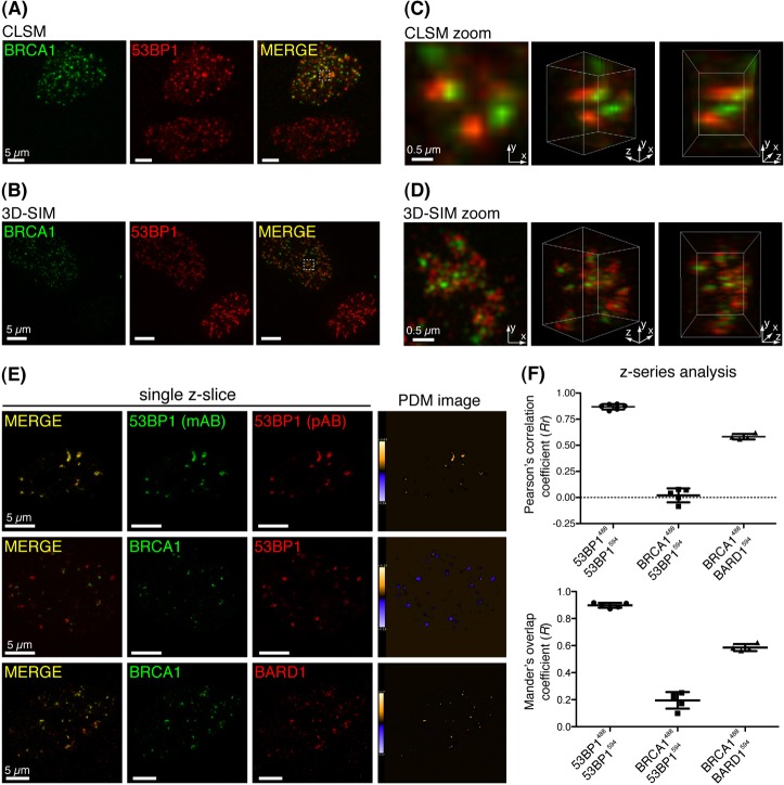
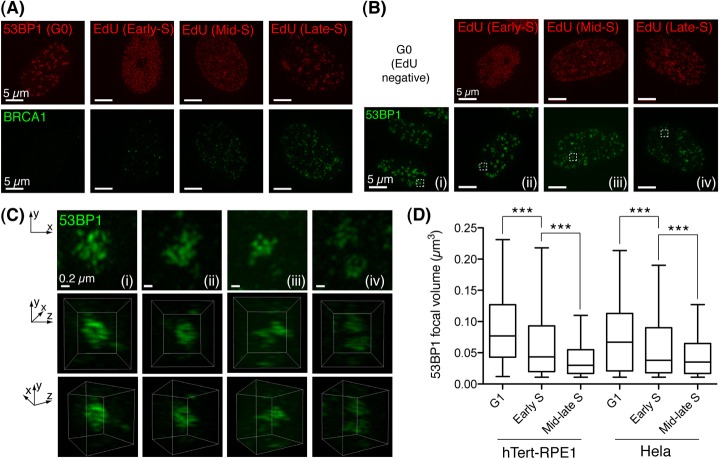
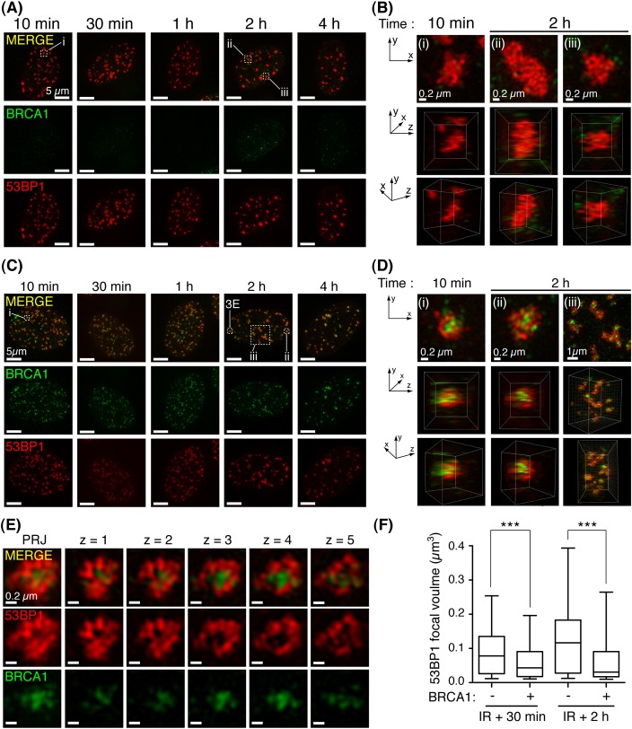
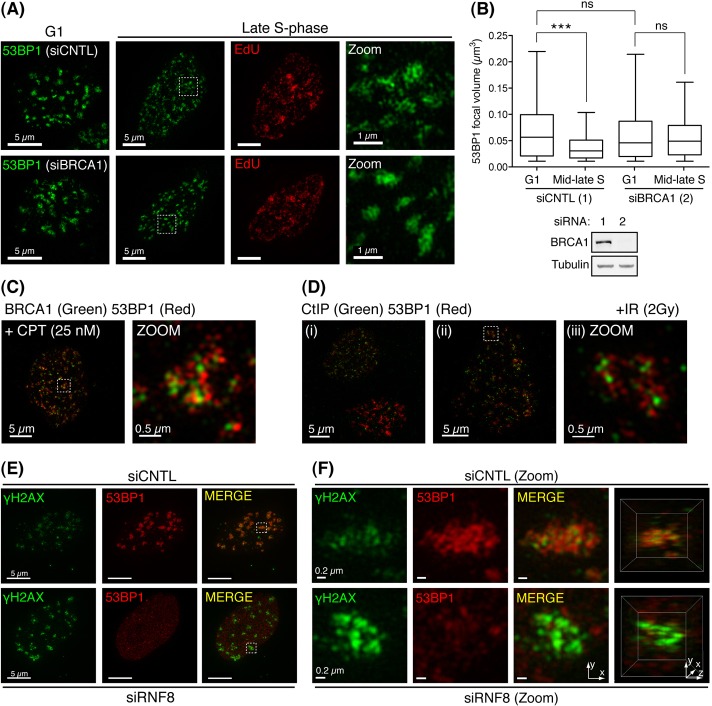
Following irradiation, numerous DNA-damage-responsive proteins rapidly redistribute into microscopically visible subnuclear aggregates, termed ionising-radiation-induced foci (IRIF). How the enrichment of proteins on damaged chromatin actually relates to DNA repair remains unclear. Here, we use super-resolution microscopy to examine the spatial distribution of BRCA1 and 53BP1 proteins within single IRIF at subdiffraction-limit resolution, yielding an unprecedented increase in detail that was not previously apparent by conventional microscopy. Consistent with a role for 53BP1 in promoting DNA double-strand break repair by non-homologous end joining, 53BP1 enrichment in IRIF is most prominent in the G0/G1 cell cycle phases, where it is enriched in dense globular structures. By contrast, as cells transition through S phase, the recruitment of BRCA1 into the core of IRIF is associated with an exclusion of 53BP1 to the focal periphery, leading to an overall reduction of 53BP1 occupancy at DNA damage sites. Our data suggest that the BRCA1-associated IRIF core corresponds to chromatin regions associated with repair by homologous recombination, and the enrichment of BRCA1 in IRIF represents a temporal switch in the DNA repair program. We propose that BRCA1 antagonises 53BP1-dependent DNA repair in S phase by inhibiting its interaction with chromatin proximal to damage sites. Furthermore, the genomic instability exhibited by BRCA1-deficient cells might result from a failure to efficiently exclude 53BP1 from such regions during S phase.


Figures

Fig. 1.
Fig. 1.
Subdiffraction-limit imaging of BRCA1 and 53BP1 in IRIF. (A) RPE1 cells recovered for 30 minutes following irradiation (2 Gy), were subjected to detergent pre-extraction and fixation before immunostaining for BRCA1 and 53BP1. Panels represent projection images constructed from multiple CLSM Z-series images. (B) Projection images constructed from 3D-SIM acquired Z-series of cells prepared exactly as in A. (C,D) Projection (left) and 3D-rendered images (other panels) were constructed from Z-series images of the indicated cell volumes (checked box) in A and B, respectively. (E) Pixel-by-pixel analysis of colocalisation of indicated fluorescently labelled proteins was performed across whole-cell 3D-SIM Z-series by intensity correlation analysis (ICA). Representative Z-slices from each dataset are presented, and the product of the differences from the mean intensity of each pixel (PDM) is shown: positive PDM pixels (an indication of co-variance) are orange, and negative PDM pixels (an indication of inverse correlation) are blue. (F) Statistical analysis of colocalisation. ICA analysis was performed across multiple Z-series (8–32 Z-slices/cell) and standard measures of image colocalisation for each labelled antibody pair are plotted. Each dot represents a single cell. Mean and s.d. are shown.
Fig. 2.
Fig. 2.
Temporal changes to BRCA1 and 53BP1 IRIF distributions. (A) RPE1 cells were G0/G1 arrested by serum starvation or asynchronous cell cultures were pulse-labelled with EdU (5 minutes, 20 µM) immediately before irradiation (2 Gy). 30 minutes later, fixed cells stained for EdU in click reactions, were counterstained for BRCA1. Representative projection images constructed from 3D-SIM-acquired Z-series datasets are shown (n≥10 per stage). (B) Cells treated exactly as in A immunostained for 53BP1. (C) Projections and 3D reconstructions of 53BP1 distribution in IRIF (top and bottom two panels, respectively) were assembled from 3D-SIM datasets of cell volumes indicated in B. (D) G1 to S-phase progression is associated with decreased 53BP1 IRIF occupancy. Volumes of isosurface rendered 53BP1 foci from multiple irradiated (harvested 1 hour following 2Gy) RPE1 (n = 32; >300 foci scored per group) and HeLa cells (n = 36; >700 foci scored per group). S-phase position was discriminated as above. EdU-negative cells with a DAPI content <S or G2 were discriminated as G1. Whiskers indicate the 5th to 95th percentile. ***P<0.0001, Mann–Whitney test.
Fig. 3.
Fig. 3.
The spatial distribution of 53BP1 and BRCA1 at DSB sites is established rapidly and persists following irradiation. (A) Irradiated cells (2 Gy), detergent pre-extracted and fixed at indicated times following irradiation, immunostained for BRCA1 and 53BP1. Representative projection images of 3D-SIM datasets (n≥8 per group). (B) Projection and 3D reconstructions of 3D-SIM datasets of cell volumes indicated in A. (C) Cells positive for co-enrichment of BRCA1 and 53BP1 in IRIF were processed as in A. (D) Projection and 3D-reconstruction images constructed from 3D-SIM datasets of cell volumes indicated in C. (E) Projection (PRJ) and single Z-slices show 53BP1 exclusion from BRCA1-associated IRIF core. (F) BRCA1 enrichment is associated with decreased 53BP1 IRIF occupancy. Volumes of isosurface-rendered 53BP1 foci from multiple irradiated cells (2 Gy) positive or negative for BRCA1 co-enrichment were analysed per time point; n = 3 per group, scoring >300 foci and >100 foci at 30 minute and 2 hour time points, respectively. Whiskers indicate the 5th to 95th percentile. ***P<0.0001, Mann–Whitney test.
Fig. 4.
Fig. 4.
BRCA1-dependent 53BP1 exclusion from IRIF coincides with S-phase DSB repair and 53BP1-associated chromatin displays reduced γH2AX. 48 hours following control (CNTL) or BRCA1 siRNA treatment, HeLa cells were processed exactly as in Fig. 2D. (A) Representative images of two independent experiments in which IRIF (>40 per cell; n≥19) were scored per experimental group. (B) Quantification of experiment shown in A (top). Whiskers indicate the 5th to 95th percentile. ns, not significant (P = 0.8639). ***P<0.0001, Mann–Whitney test. Western blot (bottom) showing BRCA1 protein depletion in CNTL and BRCA1 siRNA-treated cells (lanes 1 and 2, respectively). (C) Cells recovered for 30 minutes following acute CPT treatment (25 nM for 10 minutes) were detergent pre-extracted and fixed before BRCA1 and 53BP1 immunostaining. Representative projection images are presented (n = 10). (D) Irradiated cells (2 Gy) processed as in C were immunostained for CtIP and 53BP1. (E) 72 hours following siRNA treatment, cells were irradiated (2 Gy), recovered (30 minutes), then processed as in B with 53BP1 and γH2AX antisera. Representative projection images are presented (n≥10 in two independent experiments). (F) Projection and 3D-reconstruction images constructed from two-channel 3D-SIM datasets of cell volumes indicated in E.

References

    1. Bekker–Jensen S., Lukas C., Kitagawa R., Melander F., Kastan M. B., Bartek J., Lukas J. (2006). Spatial organization of the mammalian genome surveillance machinery in response to DNA strand breaks. J. Cell Biol. 173, 195–206 10.1083/jcb.200510130 - DOI - PMC - PubMed
    1. Bothmer A., Robbiani D. F., Feldhahn N., Gazumyan A., Nussenzweig A., Nussenzweig M. C. (2010). 53BP1 regulates DNA resection and the choice between classical and alternative end joining during class switch recombination. J. Exp. Med. 207, 855–865 10.1084/jem.20100244 - DOI - PMC - PubMed
    1. Bothmer A., Robbiani D. F., Di Virgilio M., Bunting S. F., Klein I. A., Feldhahn N., Barlow J., Chen H. T., Bosque D., Callen E., et al. (2011). Regulation of DNA end joining, resection, and immunoglobulin class switch recombination by 53BP1. Mol. Cell 42, 319–329 10.1016/j.molcel.2011.03.019 - DOI - PMC - PubMed
    1. Bunting S. F., Callén E., Wong N., Chen H. T., Polato F., Gunn A., Bothmer A., Feldhahn N., Fernandez–Capetillo O., Cao L., et al. (2010). 53BP1 inhibits homologous recombination in Brca1-deficient cells by blocking resection of DNA breaks. Cell 141, 243–254 10.1016/j.cell.2010.03.012 - DOI - PMC - PubMed
    1. Dimitrova D. S., Berezney R. (2002). The spatio-temporal organization of DNA replication sites is identical in primary, immortalized and transformed mammalian cells. J. Cell Sci. 115, 4037–4051 10.1242/jcs.00087 - DOI - PubMed

Publication types

MeSH terms

Substances