. 2012 Sep;85(1017):1226-35.
doi: 10.1259/bjr/54532316. Epub 2012 May 2.

The reversed halo sign: update and differential diagnosis

Affiliations

The reversed halo sign: update and differential diagnosis

M C B Godoy et al. Br J Radiol. 2012 Sep.

Abstract

The reversed halo sign is characterised by a central ground-glass opacity surrounded by denser air-space consolidation in the shape of a crescent or a ring. It was first described on high-resolution CT as being specific for cryptogenic organising pneumonia. Since then, the reversed halo sign has been reported in association with a wide range of pulmonary diseases, including invasive pulmonary fungal infections, paracoccidioidomycosis, pneumocystis pneumonia, tuberculosis, community-acquired pneumonia, lymphomatoid granulomatosis, Wegener granulomatosis, lipoid pneumonia and sarcoidosis. It is also seen in pulmonary neoplasms and infarction, and following radiation therapy and radiofrequency ablation of pulmonary malignancies. In this article, we present the spectrum of neoplastic and non-neoplastic diseases that may show the reversed halo sign and offer helpful clues for assisting in the differential diagnosis. By integrating the patient's clinical history with the presence of the reversed halo sign and other accompanying radiological findings, the radiologist should be able to narrow the differential diagnosis substantially, and may be able to provide a presumptive final diagnosis, which may obviate the need for biopsy in selected cases, especially in the immunosuppressed population.


Figures

Figure 1
Figure 1
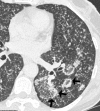
Pulmonary zygomycosis in a 22-year-old male with precursor B-cell acute lymphocytic leukaemia. (a) CT image shows the reversed halo sign (RHS) in the left upper lobe (arrows). (b) CT scan performed 2 months later shows interval development of cavitation (air crescent sign). The presence of RHS in an immunosuppressed patient is highly suggestive of pulmonary zygomycosis, especially if the patient is receiving prophylaxis for aspergillosis.
Figure 2
Figure 2
Invasive pulmonary aspergillosis in a 54-year-old female with multiple myeloma and secondary plasma cell leukaemia who had undergone chemotherapy. (a) High-resolution CT image at the lung base shows a right lower-lobe pulmonary nodule (arrow) and the reversed halo sign (RHS) in the left lower lobe (curved arrow) with adjacent ground-glass opacities. (b) CT image with mediastinal window settings shows the peripheral consolidation of the RHS (asterisks). Note a small right pleural effusion.
Figure 3
Figure 3
Paracoccidioidomycosis in a 49-year-old male, resident of a rural area in Brazil. CT image shows reversed halo sign (RHS) lesions (arrows) and bilateral small poorly marginated pulmonary nodules. The presence of RHS in a patient from an endemic region for fungal infection should raise concern for that specific fungal infection.
Figure 4
Figure 4
Tuberculosis in a 59-year-old female. High-resolution CT image shows bilateral small centrilobular nodules, tree-in-bud opacities and areas of reversed halo sign (RHS). Note the nodular appearance of the ring of consolidation of the RHS lesions (arrows), which may be helpful in the differentiation between active granulomatous disease and cryptogenic organising pneumonia. The presence of associated centrilobular nodules and tree-in-bud opacities should raise suspicion of tuberculosis.
Figure 5
Figure 5
Organising pneumonia in a 53-year-old male with graft-vs-host disease after stem cell transplantion for treatment of acute myelogenous leukaemia. CT scan shows bilateral peripheral and peribronchovascular consolidative opacities with the reversed halo sign (arrows). Open-lung biopsy showed organising pneumonia. The patient was treated with steroids with resolution of the lesions.
Figure 6
Figure 6
Sarcoidosis in a 44-year-old female. High-resolution CT image shows bilateral nodular opacities with the reversed halo sign (RHS) (arrows) and small pulmonary nodules with predominant perilymphatic distribution. The presence of the RHS associated with nodules in perilymphatic distribution and mediastinal and hilar lymphadenopathy (not shown) should raise suspicion of sarcoidosis.
Figure 7
Figure 7
Acute pulmonary embolism with pulmonary infarction in a 64-year-old female. (a) CT image shows the reversed halo sign in the superior segment of the right lower lobe. (b) CT image (mediastinal window settings) shows a clot in the distal right main pulmonary artery extending to the superior segmental branch of the right lower lobe pulmonary artery. (c) 2 month follow-up CT scan shows cavitation of the infarcted pulmonary parenchyma.
Figure 8
Figure 8
Multifocal pulmonary adenocarcinoma in a 70-year-old female. CT image shows bilateral pulmonary nodules with the reversed halo sign (black arrows) and ground-glass pulmonary nodules (white arrows).
Figure 9
Figure 9
Metastatic renal cell carcinoma in a 73-year-old male. The CT scan shows multiple bilateral lesions with reversed halo sign (RHS), biopsy proven to represent metastatic renal cell carcinoma in a background of fibrosis and necrosis. In patients with a known primary malignancy, RHS lesions may represent atypical presentation of metastatic disease. The main differential diagnosis is organising pneumonia, which can be related to drug toxicity if the patient is receiving chemotherapy.
Figure 10
Figure 10
Reversed halo sign (RHS) following radiofrequency ablation (RFA) of a pulmonary adenocarcinoma in a 64-year-old female. (a) CT image shows the adenocarcinoma in the left lower lobe. (b) CT scan performed 1 month after RFA of the tumour shows development of the RHS. When present shortly after RFA, the RHS should not be confused with recurrence of disease.
Figure 11
Figure 11
Reversed halo sign (RHS) following proton radiation therapy for a poorly differentiated squamous cell carcinoma in a 71-year-old male. (a) CT image shows the primary tumour in the left peri-hilar region. (b) CT scan performed 4 months after radiation therapy shows development of the RHS in the region of the tumour consistent with necrosis.
Figure 12
Figure 12
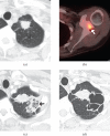
Reversed halo sign (RHS) following radiation therapy for a metastatic non-small cell lung cancer in a 59-year-old female. (a) CT image shows the primary tumour in the left upper lobe. (b) Positron emission tomography CT axial fusion image shows fludeoxyglucose (FDG) avid metastasis in the left scapula (arrow). (c) CT scan 1 month after chemotherapy and palliative radiation therapy to the left scapula shows development of the RHS in the left lung apex (arrow) adjacent to the primary tumour, consistent with radiation pneumonitis. (d) A follow-up CT scan performed 4 months after (c) shows development of cavitation in the region of the RHS. When present within the radiation field shortly after radiation therapy, the RHS should not be confused with infection or recurrence or disease.
Figure 13
Figure 13
Differential diagnosis of the reversed halo sign (RHS). AIA, angioinvasive aspergillosis; AIS, adenocarcinoma in situ; COP, cryptogenic organising pneumonia; LPA, lepidic predominant adenocarcinoma; N, no; NSIP, non-specific interstitial pneumonia; OP, organising pneumonia; PJP, Pneumocystis jiroveci pneumonia; PZ, pulmonary zygomycosis; RFA, radiofrequency ablation; Y, yes. *Includes immunosuppression, close contact with a person with tuberculosis, residence in or immigration from country where tuberculosis is common, residence in nursing home or prison, homelessness. **Includes cavitary lesions, lobular consolidation, centrilobular nodules and tree-in-bud opacities, as well as nodularity of the wall of the RHS and small nodules inside the RHS. ***Suspected in patients with acquired immunedeficiency syndrome. +Upper respiratory tract disease and non-specific glomerulonephritis, and elevation of cytoplasmic antineutrophil cytoplasmic antibody titres. ++Nodularity of the wall of the RHS and small nodules inside the RHS, possibly associated with symmetrical bilateral hilar and right paratracheal lymphadenopathy and small nodules with perilymphatic distribution. Associated with connective tissue diseases, aspiration, drug toxicity, hypersensitivity pneumonitis, infection or radiation therapy. ††Less likely.

References

    1. Hansell DM, Bankier AA, MacMahon H, McLoud TC, Muller NL, Remy J. Fleischner Society: glossary of terms for thoracic imaging. Radiology 2008;246:697–722 - PubMed
    1. Zompatori M, Poletti V, Battista G, Diegoli M. Bronchiolitis obliterans with organizing pneumonia (BOOP), presenting as a ring-shaped opacity at HRCT (the atoll sign). A case report. Radiol Med 1999;97:308–10 - PubMed
    1. Voloudaki AE, Bouros DE, Froudarakis ME, Datseris GE, Apostolaki EG, Gourtsoyiannis NC. Crescentic and ring-shaped opacities. CT features in two cases of bronchiolitis obliterans organizing pneumonia (BOOP). Acta Radiol 1996;37:889–92 - PubMed
    1. Tzilas V, BA, Provata A, Koti A, Tzouda V, Tsoukalas G. The “reversed halo” sign in pneumonococcal pneumonia: a review with a case report. Eur Rev Med Pharmacol Sci 2010;14:481–86 - PubMed
    1. Marchiori E, Zanetti G, Escuissato DL, Souza AS, Jr, Meirelles GD, Fagundes J, et al. Reversed halo sign: high-resolution CT findings in 79 patients. Chest 2012;141:1260–6 - PubMed

MeSH terms