. 2012 Jul;86(13):7360-71.
doi: 10.1128/JVI.00157-12. Epub 2012 May 2.

Evidence for a genetic and physical interaction between nonstructural proteins NS1 and NS4B that modulates replication of West Nile virus

Affiliations

Evidence for a genetic and physical interaction between nonstructural proteins NS1 and NS4B that modulates replication of West Nile virus

Soonjeon Youn et al. J Virol. 2012 Jul.

Abstract

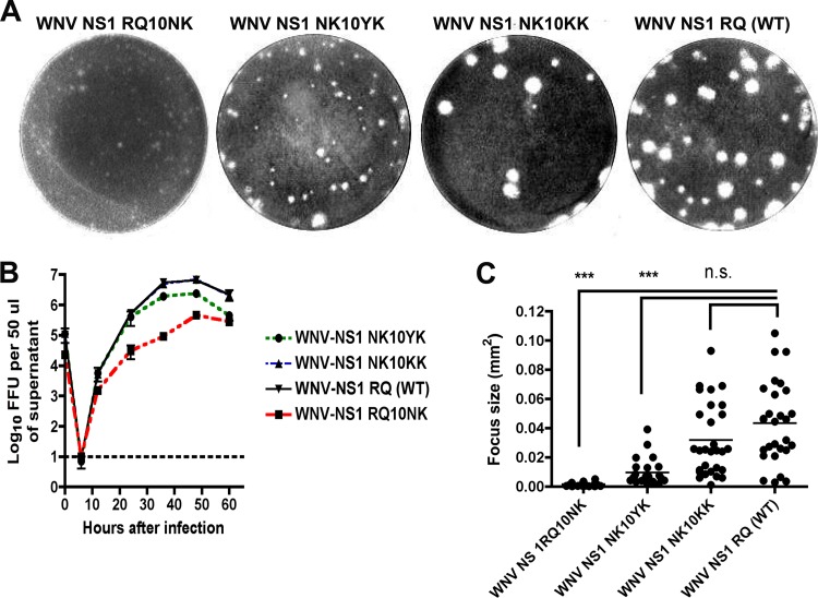
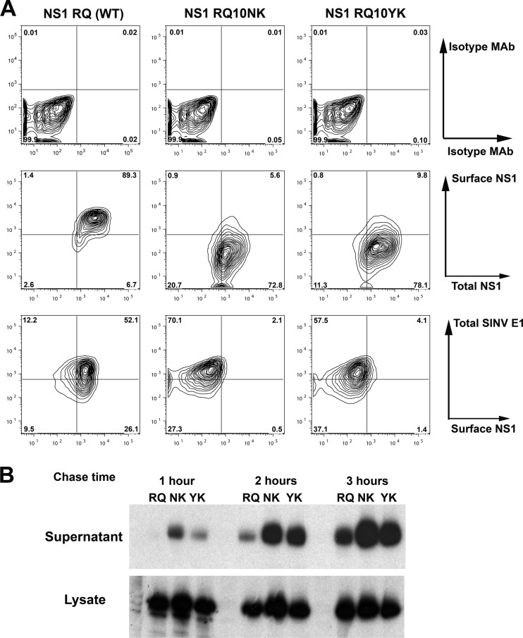
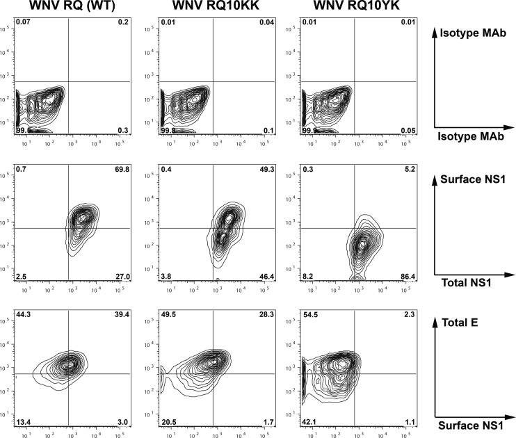
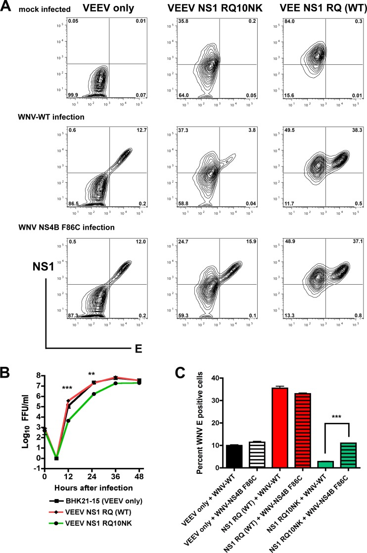
Flavivirus NS1 is a nonstructural glycoprotein that is expressed on the cell surface and secreted into the extracellular space. Despite its transit through the secretory pathway, NS1 is an essential gene linked to early viral RNA replication. How this occurs has remained a mystery given the disparate localization of NS1 and the viral RNA replication complex, as the latter is present on the cytosolic face of the endoplasmic reticulum (ER). We recently identified an N-terminal di-amino acid motif in NS1 that modulates protein targeting and affected viral replication. Exchange of two amino acids at positions 10 and 11 from dengue virus (DENV) into West Nile virus (WNV) NS1 (RQ10NK) changed its relative surface expression and secretion and attenuated infectivity. However, the phenotype of WNV containing NS1 RQ10NK was unstable, as within two passages heterogeneous plaque variants were observed. Here, using a mutant WNV encoding the NS1 RQ10NK mutation, we identified a suppressor mutation (F86C) in NS4B, a virally encoded transmembrane protein with loops on both the luminal and cytoplasmic sides of the ER membrane. Introduction of NS4B F86C specifically rescued RNA replication of mutant WNV but did not affect the wild-type virus. Mass spectrometry and coimmunoprecipitation studies established a novel physical interaction between NS1 and NS4B, suggesting a mechanism for how luminal NS1 conveys signals to the cytoplasm to regulate RNA replication.


Figures

Fig 1
Fig 1
Comparison of plaque size and growth kinetics of WNV NS1 RQ10NK revertants. (A) WNV NS1 RQ10NK (left) was serially passaged in BHK21-15 cells. Within a few passages emerged a more rapidly growing small-plaque variant (WNV NS1 NK10YK, second left) and a large-plaque variant (WNV NS1 NK10KK, second right), which had a plaque size similar to that of the wild-type virus (WNV NS1 RQ, right). Sequences were generated after plaque purification and were representative of multiple isolates from each group. (B) Single-step growth kinetics of WNV NS1 RQ10NK and revertant viruses. WNV NS1 RQ10NK, WNV NS1 NK10YK, WNV NS1 NK10KK, and WNV NS1 RQ (WT) were added to BHK21-15 cells at an MOI of 1. At each of the indicated time points, virus in the supernatant was titrated by focus-forming assay. The data are the representative results of three independent experiments performed in triplicate. The growth of WNV NS1 NK10YK, WNV NS1 NK10KK, and WNV NS1 RQ (WT) was enhanced compared to that of WNV NS1 RQ10NK at 12, 24, 36, and 48 h (P < 0.05). Note that the growth curves for WNV NS1 NK10KK and WNV NS1 RQ (WT) are quite similar and difficult to distinguish on this graph. (C) Focus size comparison of WNV NS1 RQ10NK, WNV NS1 NK10YK, WNV NS1 NK10KK, and WNV NS1 RQ (WT). The data were obtained from several independent wells and were analyzed using a Biospot counter (Cellular Technology). Asterisks indicate statistical significance (***, P < 0.0001); n.s., not statistically significant. As described in Results, the WNV NS1 NK10YK revertant virus also contained a second site mutation in NS4B (F86C).
Fig 2
Fig 2
Ectopically expressed NS1 RQ10YK showed cellular targeting similar to that of NS1 RQ10NK. (A) BHK21-15 cells were infected with recombinant SINV (MOI of 10) ectopically expressing NS1 RQ (wild type), NS1 RQ10NK, or NS1 RQ10YK. At 14 h after infection, surface NS1 and total NS1 were measured by flow cytometry (middle panels) after sequentially incubating intact and permeabilized cells with different anti-NS1 MAbs (see Materials and Methods). Isotype controls also were performed (top panels). As an infectivity control, total SINV E1 expression level was measured using a polyclonal anti-E1 antibody (bottom panels). Contour plots are shown and are representative of three independent experiments. (B) Biosynthesis of wild-type and mutant NS1. BHK21-15 cells were infected at an MOI of 5 with SINV that ectopically expressed NS1 RQ (wild type), NS1 RQ10NK, or NS1 RQ10YK. At 15 h after infection, cells were placed in cysteine-methionine-free medium for 20 min, pulsed with [35S]cysteine-methionine, and then chased for 1, 2, or 3 h. Cell supernatants or lysates were harvested, immunoprecipitated with 4NS1 MAb and protein A-Sepharose beads, and electrophoresed. The data are the representative results of two independent experiments.
Fig 3
Fig 3
Cellular targeting of NS1 in the context of infection by revertant WNV. BHK21-15 cells were infected at an MOI of 1 with WNV NS1 RQ (wild type) or the revertants (WNV NS1 RQ10KK or WNV NS1 RQ10YK) generated by passage (Fig. 1), and the cellular targeting of NS1 was analyzed. At 30 h after infection, surface and total NS1 levels were measured by flow cytometry (middle panels) after sequentially incubating intact and permeabilized cells with different anti-NS1 MAbs. Isotype controls also were performed (top panels). As an infectivity control, total WNV E expression level was measured using a specific MAb (WNV E16) (bottom panels). Contour plots are shown and are representative of results from three independent experiments.
Fig 4
Fig 4
The F86C mutation in NS4B is specifically required for growth of WNV with mutant NS1. (A) Single-step growth kinetics of WNV NS1 RQ (wild type) and WNV NS1 RQ + NS4B F86C were compared. All viruses were generated from a two-plasmid infectious clone of WNV as detailed in Materials and Methods. BHK21-15 cells were infected at an MOI of 5, and at the indicated time points, virus was harvested from supernatants for titration by focus-forming assay. The data are the representative results of three independent experiments performed in triplicate. (B) Single-step growth kinetics of WNV containing NS1 RQ10NK plus NS4B WT, NS1 RQ10NK plus NS4B F86C, NS1 RQ10YK plus NS4B WT, NS1 RQ10YK plus NS4B F86C, and the parent NS1 RQ (WT) plus NS4B WT. All viruses were generated from an infectious clone of WNV and infected and titrated as described for panel A. The data are representative of results of three independent experiments performed in triplicate, and viruses containing NS1 RQ10NK were statistically different at several time points after infection. (C) Focus size comparison of WNV engineered to have NS1 RQ10YK plus NS4B WT or NS1 RQ10YK plus NS4B F86C. The data are pooled from several independent experiments, and differences were statistically significant (***, P < 0.0001) (D) Amino acid sequence alignment after sequencing of viral RNA harvested from the indicated P0 stocks that were generated after transfection into BHK21-15 cells of in vitro transcribed WNV RNA. Adaptive or stabilizing mutations are explicitly identified. Note that no adventitious mutations were observed in the template cDNA or the in vitro transcribed RNA prior to electroporation.
Fig 5
Fig 5
Ectopically expressed NS1 RQ10NK acts as a dominant negative for infection of WNV-WT but not WNV containing the NS4B F86C suppressor mutation. (A) BHK21-15 cells propagating VEEV subgenomic replicons encoding NS1 with an N-terminal stop codon (VEEV only), NS1 RQ10NK, or NS1 RQ (wild type) were mock infected (top panels) or superinfected at an MOI of 1 with WNV-WT (middle panels) or WNV containing the NS4B F86C suppressor mutation (WNV NS4B F86C, bottom panels), both of which were generated from the infectious cDNA clone. At 12 h after infection, cells were fixed and permeabilized, and total levels of NS1 and E protein expression were assessed by flow cytometry after immunostaining with 4NS1 or humanized WNV-E16 MAbs. (B) Viral growth curve analysis. BHK21-15 cells (mock infected) or BHK21-15 cells propagating VEEV subgenomic replicons as listed above were mock infected or superinfected at an MOI of 10 with WNV-WT or WNV NS4B F86C. At the indicated time points, virus was harvested from the supernatant and titrated by focus-forming assay. The data are the average of three independent experiments performed in duplicate, and the asterisks indicate statistically significant differences between viruses grown in cells expressing NS1 RQ10NK and NS1 RQ (wild type). (C) Summary of E protein expression by flow cytometry after infection of WNV-WT or WNV-NS4B F86C in cells propagating VEEV replicons with an N-terminal stop codon of NS1 (VEEV only), NS1 RQ10NK, or NS1 RQ (wild type). Results are averaged from three independent experiments, error bars denote standard error of the mean, and asterisks indicate statistically significant differences (***, P < 0.0001).
Fig 6
Fig 6
Dominant negative effect of NS1 RQ10NK mutation on WNV RNA replication is rescued by the NS4B F86C suppressor mutation. BHK21-15 cells propagating VEEV replicon expressing NS1 RQ (wild type) or NS1 RQ10NK NS1 were superinfected with WNV-WT (A) or WNV NS4B F86C (B) at an MOI of 10. At 3, 6, and 12 h after infection, total RNA was extracted from cells and WNV RNA was measured by real-time quantitative RT-PCR and compared to 18S rRNA. A standard curve of 18S rRNA per BHK21-15 cell was used for normalization. The data are the average of results from three independent experiments performed in duplicate, and asterisks indicate statistically significant differences (**, P < 0.01; ***, P < 0.0001).
Fig 7
Fig 7
Oligomeric forms of soluble WT and RQ10NK NS1 after purification. WT and RQ10NK NS1 were purified by sequential antibody affinity (9NS1 Sepharose) and size exclusion (Superdex-200) chromatography from supernatants of BHK21-15 cell lines propagating a VEEV replicon that ectopically expressed the WT and variant NS1 proteins. Fractions corresponding to the peaks of the size exclusion chromatography were subjected to SDS-PAGE and silver staining (A) or Western blotting (B) with 10NS1 MAb. Fractions A7 and A8, higher-order NS1 oligomers (WT only); B2 and B3, hexamer (WT only); B8 to B10, tetrameric form; and C4 and C5, dimer. (C) Based on a calibration curve (standards ranging from 13.7 to 669 kDa), the approximate molecular sizes were determined and are indicated in the chromatography tracings.
Fig 8
Fig 8
Physical interaction between NS1 and NS4B. (A) Coimmunoprecipitation and Western blotting. BHK21-15 cells were mock infected or infected with WNV-WT at an MOI of 10. At 28 h after infection, cells were lysed and NS1 proteins were immunoprecipitated with anti-NS1 MAb and protein A-Sepharose. After extensive washing, beads were boiled in SDS sample buffer, and the eluate was electrophoresed and subjected to Western blotting with a polyclonal anti-NS4B antibody. (B to D) Mass spectrometry (MS) identifies an interaction between WNV NS1 and NS4B. (B) Workflow for the immunoaffinity isolation of NS1 protein complexes. (C) Summary of viral proteins identified by mass spectrometry (nLC-MS/MS using an LTQ Orbitrap Velos) as coisolated with NS1 in WNV-KUNV-infected cells. The number of unique peptides and identifications of boundary peptides that indicate mature forms are shown. Three viral proteins coisolated with NS1 and validated as mature proteins are shown in bold. M, molecular mass. (D) Representative collision-induced dissociation (CID) MS/MS spectrum of a tryptic peptide of NS4B demonstrating its association with WNV NS1.

Similar articles

Cited by

References

    1. Alcon-LePoder S, et al. 2005. The secreted form of dengue virus nonstructural protein NS1 is endocytosed by hepatocytes and accumulates in late endosomes: implications for viral infectivity. J. Virol. 79:11403–11411 - PMC - PubMed
    1. Ambrose RL, Mackenzie JM. 2011. West Nile virus differentially modulates the unfolded protein response to facilitate replication and immune evasion. J. Virol. 85:2723–2732 - PMC - PubMed
    1. Avirutnan P, et al. 2010. Antagonism of the complement component C4 by flavivirus non-structural protein NS1. J. Exp. Med. 207:793–806 - PMC - PubMed
    1. Avirutnan P, et al. 2011. Binding of flavivirus nonstructural protein NS1 to C4b binding protein modulates complement activation. J. Immunol. 187:424–433 - PMC - PubMed
    1. Avirutnan P, et al. 2007. Secreted NS1 of dengue virus attaches to the surface of cells via interactions with heparan sulfate and chondroitin sulfate E. PLoS Pathog. 3:e183 doi:10.1371/journal.ppat.0030183 - DOI - PMC - PubMed

Publication types

Substances

LinkOut - more resources