. 2012 Jul;86(14):7554-64.
doi: 10.1128/JVI.00212-12. Epub 2012 May 2.

An in-frame deletion in the NS protein-coding sequence of parvovirus H-1PV efficiently stimulates export and infectivity of progeny virions

Affiliations

An in-frame deletion in the NS protein-coding sequence of parvovirus H-1PV efficiently stimulates export and infectivity of progeny virions

Nadine Weiss et al. J Virol. 2012 Jul.

Abstract

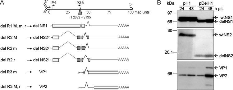
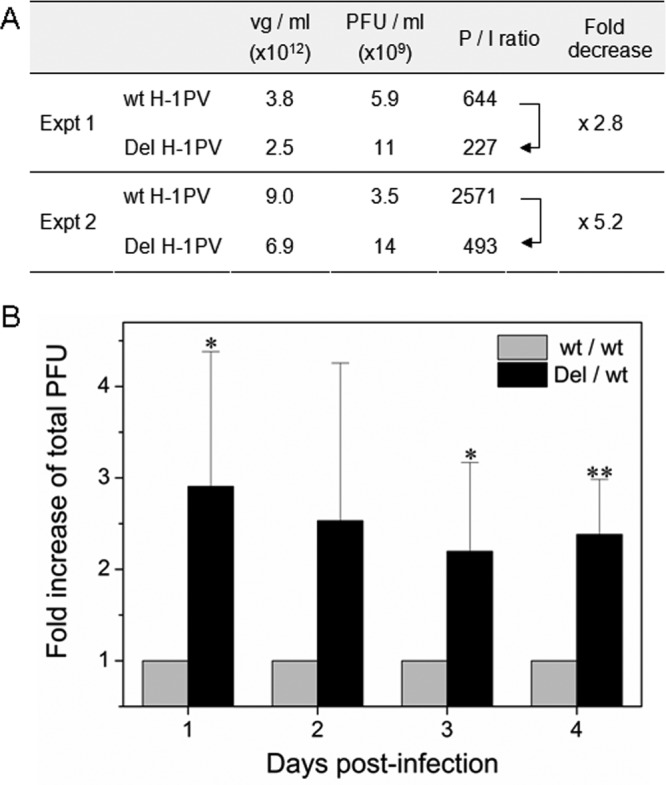
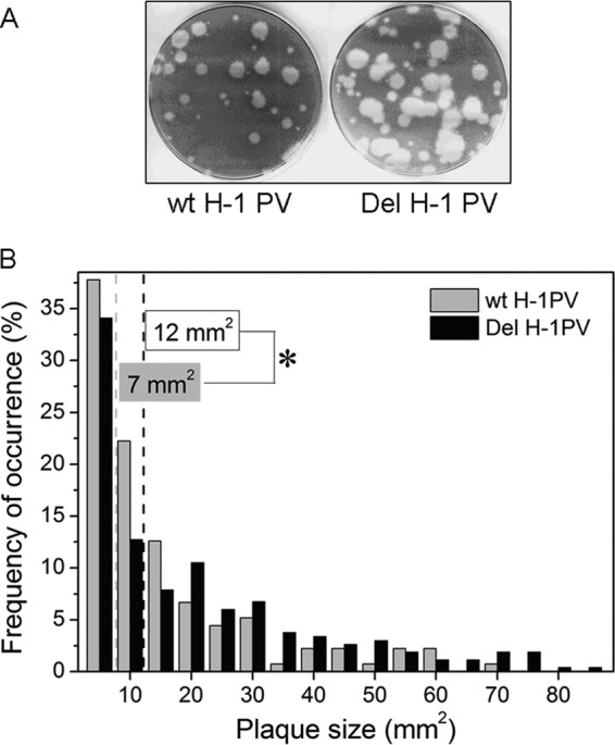
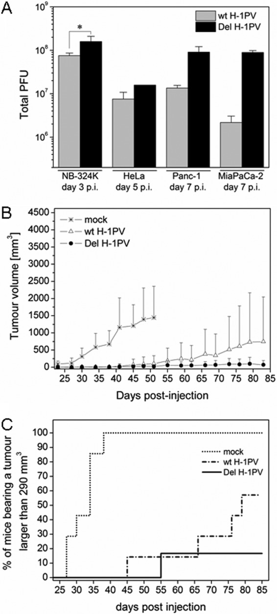
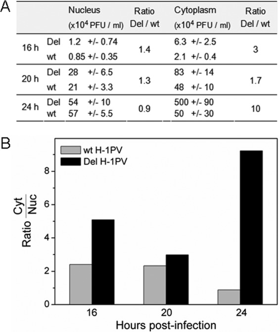
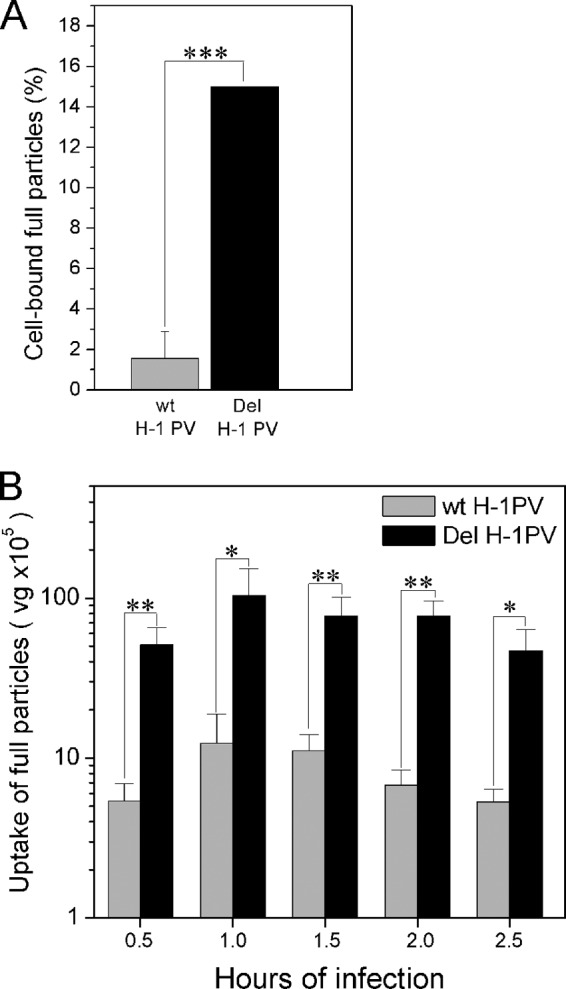
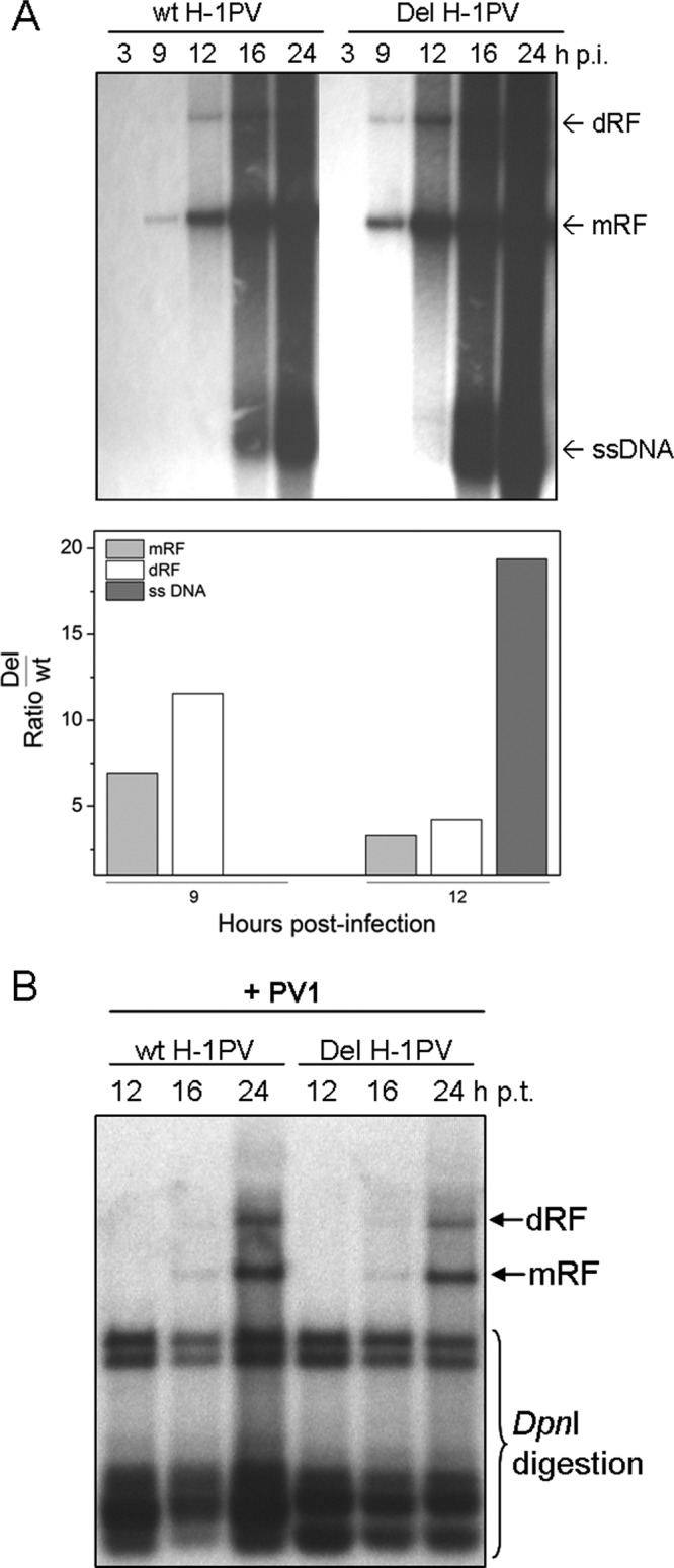
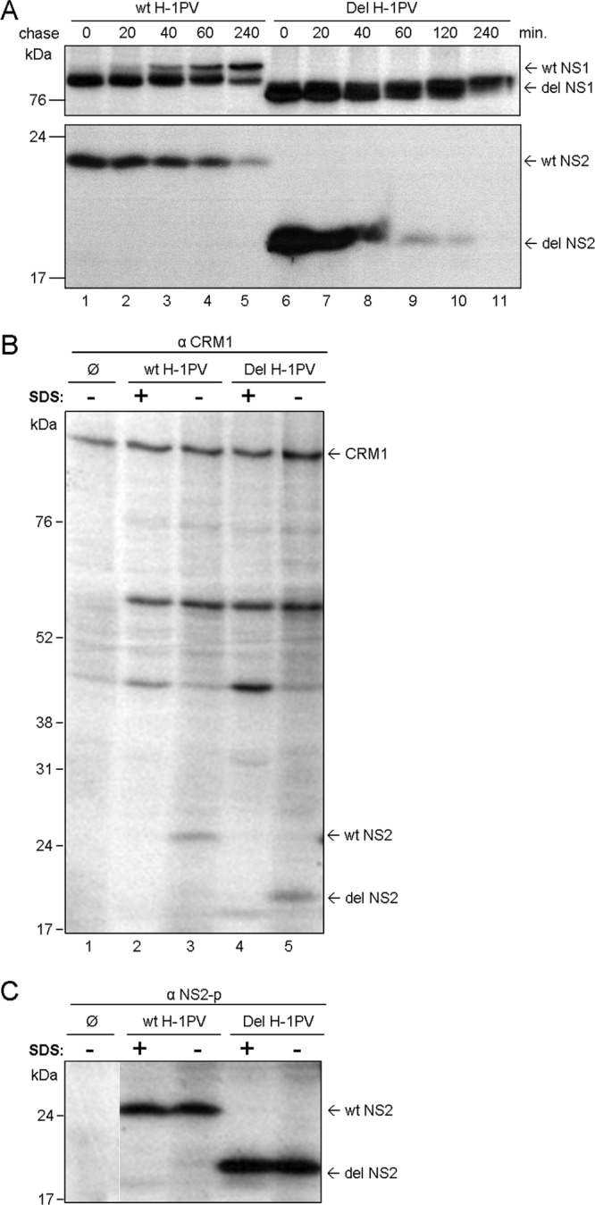
An in-frame, 114-nucleotide-long deletion that affects the NS-coding sequence was created in the infectious molecular clone of the standard parvovirus H-1PV, thereby generating Del H-1PV. The plasmid was transfected and further propagated in permissive human cell lines in order to analyze the effects of the deletion on virus fitness. Our results show key benefits of this deletion, as Del H-1PV proved to exhibit (i) higher infectivity (lower particle-to-infectivity ratio) in vitro and (ii) enhanced tumor growth suppression in vivo compared to wild-type H-1PV. This increased infectivity correlated with an accelerated egress of Del H-1PV progeny virions in producer cells and with an overall stimulation of the viral life cycle in subsequently infected cells. Indeed, virus adsorption and internalization were significantly improved with Del H-1PV, which may account for the earlier appearance of viral DNA replicative forms that was observed with Del H-1PV than wild-type H-1PV. We hypothesize that the internal deletion within the NS2 and/or NS1 protein expressed by Del H-1PV results in the stimulation of some step(s) of the viral life cycle, in particular, a maturation step(s), leading to more efficient nuclear export of infectious viral particles and increased fitness of the virus produced.


Figures

Fig 1
Fig 1
Schematic representation of Del H-1PV genomic organization and accumulation of the nonstructural delNS1 and delNS2 and capsid VP1 and VP2 proteins. (A) Schematic representation of the viral genome (top) with the 3′ and 5′ palindromic regions and the P4 and P38 promoters. The triangle indicates the position of the deletion that was introduced in the H-1PV genome. (Bottom) Scheme of the shortened transcripts (lines) and proteins (boxes). The hatched region indicates the localization of the deletion. (B) Accumulation of the nonstructural and capsid proteins expressed from pDelH1 and pH1 in 293T cells. Protein extracts were prepared at 24 h and 48 h posttransfection with either pH1 or pDelH1 and subjected to 12% (NS) or 10% (VP) SDS-PAGE followed by immunoblotting using polyclonal rabbit antisera recognizing either the NS1 protein (top), the NS2P protein (middle), or the VP proteins (bottom).
Fig 2
Fig 2
Del H-1PV progeny production and infectivity are increased in permissive human NB-324K cells. (A) Increased infectivity of Del H-1PV versus wt H-1PV particles. Cells infected with Del H-1PV or wt H-1PV (MOI, 0.003 PFU) were harvested at day 3 p.i. Titers of infectious and full particles were determined by plaque assay (given in PFU/ml) and qPCR (given in vg/ml), respectively. The particle-to-infectivity (P/I) ratio and the fold decrease of the P/I ratio between wt H-1PV and Del H-1PV are indicated. (B) Time course production of Del H-1PV and wt H-1PV. Cells were infected at an MOI of 0.5 PFU. The amounts of infectious virus were determined by plaque assay (infectious particles from both cell extract and culture medium; total PFU). Data are shown as fold increase of Del H-1PV progeny production over wt H-1PV progeny production and represent the means of three independent experiments with the standard deviation (SD). *, P < 0.05; **, P < 0.01.
Fig 3
Fig 3
Enhanced propagation of Del H-1PV compared to wild-type H-1PV in infected NB-324K cells. (A) Illustration of plaque sizes produced by wt H-1PV and Del H-1PV after infection of NB-324K cells. (B) Plaque analysis using the Java-based image-processing software ImageJ. Plaque sizes are given in mm2, and frequencies of occurrence are expressed as a percentage of the total number of analyzed plaques: n = 135 for wt H-1PV and n = 267 for Del H-1PV. The means of the plaque sizes produced by wt H-1PV or Del H-1PV are indicated with dotted gray and black lines, with their values given in gray and black boxes, respectively. *, P < 0.05.
Fig 4
Fig 4
Del H-1PV production and ability for tumor suppression are more efficient than those for wt H-1PV. (A) Del H-1PV progeny production is increased in various human cell lines. Infectious progeny production of Del H-1PV and wt H-1PV was measured after infection (MOI, 0.5 PFU/cell) of human NB-324K, HeLa, and PDAC Panc-1 and MiaPaCa-2 cell lines. The yields of infectious particles (total numbers of PFU) recovered from both cell supernatants and pellets were determined by plaque assay at days 3, 5, and 7 p.i. for NB-324K, HeLa, and both PDAC cell lines, respectively. *, P < 0.05. (B and C) Del H-1PV is more efficient in suppressing the growth of Panc-1 cell-derived tumors than wt H-1PV. Panc-1 cells (5 × 10⁶) were buffer treated (mock) or infected with wt H-1PV or Del H-1PV at an MOI of 1 RU/cell and subcutaneously implanted into the right flank of BALB/c nude mice. Tumor appearance and development were monitored over time. (B) Tumor volumes are given as means for 6 or 7 mice/group (± SD); (C) the percentage of mice bearing a tumor larger than 290 mm3 is plotted as a function of time postimplantation.
Fig 5
Fig 5
Earlier nuclear release of Del H-1PV progeny virions in NB-324K cells. NB-324K cells were infected with Del H-1PV or wt H-1PV at an MOI of 1 PFU/cell and further incubated in the presence of neutralizing antibodies. Cells were harvested at the indicated points of time p.i. and subjected to a nuclear-cytoplasmic fractionation. Infectious virus particles present in both fractions were titrated by plaque assay. (A) Titers (in PFU/ml ± SD) and ratio of Del H-1PV to wt H-1PV infectious particles are given for nuclear and cytoplasmic fractions. (B) Virus distribution between cytoplasmic (Cyt) and nuclear (Nuc) fractions (cytoplasmic fraction/nuclear fraction ratio) of Del H-1PV and wt H-1PV.
Fig 6
Fig 6
Improved cell binding (A) and uptake (B) of Del H-1PV particles compared with wt H-1PV particles. (A) NB-324K cells were infected with either wt H-1PV or Del H-1PV at an MOI of 104 vg/cell and 4°C for 1 h. The amount of cell-bound full particles was determined by qPCR and expressed as the percentage of input viral genome. ***, P < 0.001. (B) NB-324K cells were infected at an MOI of 104 vg/cell and 37°C. Cells were recovered at the indicated times of infection and treated with trypsin-EDTA to remove cell-bound particles. Following cell lysis, the amount of internalized full particles was determined by qPCR and given in vg. *, P < 0.05; **, P < 0.01.
Fig 7
Fig 7
Increased accumulation of Del H-1PV DNA replicative forms in infected—but not transfected—NB-324K cells. (A) NB-324K cells (9 × 10⁵) were infected with wt H-1PV or Del H-1PV at an MOI of 6,844 vg/cell. At 2 h postinfection, neutralizing antibodies were added and cells were harvested at the indicated points of time p.i. Viral DNA replicative forms purified from cell lysates were separated by agarose gel electrophoresis and subjected to Southern blotting. Signal intensities of Del H-1PV and wt H-1PV replicative forms (mRF and dRF) and ssDNA were quantified by PhosphorImager analysis. (Top) Viral replicative intermediates revealed through hybridization with an NS1-specific 32P-labeled DNA probe; (bottom) ratio of signal intensities corresponding to Del H-1PV replicative forms and ssDNA over those corresponding to wt H-1PV ones. (B) NB-324K cells (2 × 10⁶) were transfected with 6 μg of pH1 or pDelH1. Neutralizing antibodies (+ PV1) were added 4 h p.t., and cells were harvested at the indicated points of time p.t. Viral DNA replicative forms purified from cell lysates were DpnI digested and analyzed by Southern blotting.
Fig 8
Fig 8
Increased neosynthesis and degradation of delNS2P compared to wt NS2P protein. (A) Synthesis and stability of delNS2P and wt NS2P in NB-324K cells. Cells were infected with Del H-1PV or wt H-1PV at an MOI of 10 PFU. Proteins were metabolically labeled with [35S]cysteine/methionine for 30 min (pulse), and cells were further incubated in nonradioactive medium (chase) prior to extraction of the proteins in RIPA buffer at the indicated times. Proteins were immunoprecipitated using rabbit polyclonal antisera recognizing either the NS1 protein (top) or the NS2P protein (bottom), separated by 12% SDS-PAGE, and visualized by autoradiography. (B and C) DelNS2P is able to interact with the nuclear export factor CRM1. Proteins were metabolically labeled with [35S]cysteine/methionine for 2 h before being extracted in the presence (lanes +) or absence (lanes −) of SDS. Proteins were (co-)immunoprecipitated using rabbit polyclonal antibodies recognizing either CRM1 (B) or NS2P (C), separated by 12% SDS-PAGE, and visualized by autoradiography. The positions of wt NS1 (83 kDa), delNS1 (∼76 kDa), wt NS2P (35 kDa), delNS2P (∼18 kDa), and CRM1 (110 kDa) are indicated. Ø, mock-treated cells.

Similar articles

Cited by

References

    1. Baer S, Daeffler L, Rommelaere J, Nueesch JPF. 2008. Vesicular egress of non-enveloped lytic parvoviruses depends on gelsolin functioning. PLoS Pathog. 4:e1000126 doi:10.1371/journal.ppat.1000126 - DOI - PMC - PubMed
    1. Ball-Goodrich LJ, Leland SE, Johnson EA, Paturzo FX, Jacoby RO. 1998. Rat parvovirus type 1: the prototype for a new rodent parvovirus serogroup. J. Virol. 72:3289–3299 - PMC - PubMed
    1. Besselsen DG, et al. 1996. Molecular characterization of newly recognized rodent parvoviruses. J. Gen. Virol. 77(Pt 5):899–911 - PubMed
    1. Besselsen DG, Romero MJ, Wagner AM, Henderson KS, Livingston RS. 2006. Identification of novel murine parvovirus strains by epidemiological analysis of naturally infected mice. J. Gen. Virol. 87:1543–1556 - PubMed
    1. Bodendorf U, Cziepluch C, Jauniaux J-C, Rommelaere J, Salome N. 1999. Nuclear export factor CRM1 interacts with nonstructural proteins NS2 from parvovirus minute virus of mice. J. Virol. 73:7769–7779 - PMC - PubMed

Publication types

Substances

LinkOut - more resources