. 2012 Jul;86(14):7508-19.
doi: 10.1128/JVI.06725-11. Epub 2012 May 2.

Recombinant canine distemper virus strain Snyder Hill expressing green or red fluorescent proteins causes meningoencephalitis in the ferret

Affiliations

Recombinant canine distemper virus strain Snyder Hill expressing green or red fluorescent proteins causes meningoencephalitis in the ferret

M Ludlow et al. J Virol. 2012 Jul.

Abstract

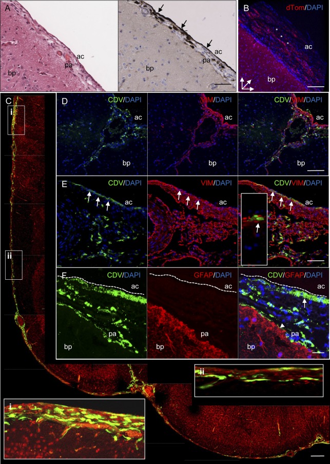
The propensity of canine distemper virus (CDV) to spread to the central nervous system is one of the primary features of distemper. Therefore, we developed a reverse genetics system based on the neurovirulent Snyder Hill (SH) strain of CDV (CDV(SH)) and show that this virus rapidly circumvents the blood-brain and blood-cerebrospinal fluid (CSF) barriers to spread into the subarachnoid space to induce dramatic viral meningoencephalitis. The use of recombinant CDV(SH) (rCDV(SH)) expressing enhanced green fluorescent protein (EGFP) or red fluorescent protein (dTomato) facilitated the sensitive pathological assessment of routes of virus spread in vivo. Infection of ferrets with these viruses led to the full spectrum of clinical signs typically associated with distemper in dogs during a rapid, fatal disease course of approximately 2 weeks. Comparison with the ferret-adapted CDV(5804P) and the prototypic wild-type CDV(R252) showed that hematogenous infection of the choroid plexus is not a significant route of virus spread into the CSF. Instead, viral spread into the subarachnoid space in rCDV(SH)-infected animals was triggered by infection of vascular endothelial cells and the hematogenous spread of virus-infected leukocytes from meningeal blood vessels into the subarachnoid space. This resulted in widespread infection of cells of the pia and arachnoid mater of the leptomeninges over large areas of the cerebral hemispheres. The ability to sensitively assess the in vivo spread of a neurovirulent strain of CDV provides a novel model system to study the mechanisms of virus spread into the CSF and the pathogenesis of acute viral meningitis.


Figures

Fig 1
Fig 1
Construction and characterization of recombinant CDVs. (A) Schematic representation of pCDVSH full-length infectious clone. A subclone containing the H-L gene boundary was produced following digestion of pCDVSH with RsrII and SwaI to facilitate the introduction of a unique PmeI site (B) as indicated by the gray shading. (C) An additional transcription unit containing EGFP or dTom was inserted at the H-L gene boundary using the unique PmeI site to produce the pCDVSHEGFP(6) and pCDVSHdTom(6) full-length clones. (D) Multistep growth curves of CDVR252, CDV5804P, CDVSH, rCDVSHdTom(6), and rCDVSHEGFP(6) in VerodogSLAM cells. Virus was harvested at 12-h intervals up to 96 h postinfection. Viral titers were determined as the number of TCID50/ml in an endpoint titration test. Measurements shown are averages of triplicates ± SE. (E) Phase-contrast photomicrographs taken at 72 h.p.i. of VerodogSLAM cells with Opti-MEM (mock), CDVR252, CDV5804P, CDVSH, rCDVSHdTom(6), and rCDVSHEGFP(6). Bar, 200 μm.
Fig 2
Fig 2
Course of disease in ferrets infected with nonrecombinant or recombinant CDV strains. (A) Body temperatures; (B) differential lymphocyte counts normalized for each animal to numbers of lymphocyte present at day 0; (C) survival curves. CDVR252, n = 6; rCDV5804P, n = 4; CDVSH, n = 3, rCDVSHdTom(6), n = 6; rCDVSHEGFP(6), n = 3. Data are shown as means per group. ***, the life expectancy of CDVR252-infected ferrets estimated by the Kaplan-Meier method was significantly greater (P < 0.05) than that of other ferret groups upon comparison using a log rank test.
Fig 3
Fig 3
Detection of macroscopic fluorescence in tissues from rCDVSHEGFP(6) (green)- or rCDVSHdTom(6) (red)-infected ferrets. (A) Extensive virus infection of mucosal surface surrounding the eye (arrow), with infection also observed in the skin beneath the mouth. No green fluorescence is observed around the mouth or nose of a CDVR252-infected ferret (inset i), in contrast to the high levels of fluorescence observed around analogous regions of an rCDVSHEGFP(6)-infected ferret (inset ii). (B) Infection of the gingiva (arrow). (C) Infection of skin epidermis in the abdominal region. (D) Numerous discrete foci of infection were visible on the surface of the tongue, with prominent infection of adjacent lymphoid tissues and tonsils (arrows). (E) Infection of salivary gland (arrow) and lymph nodes (arrowheads). (F) Multiple large foci of infection were observed throughout all lobes of the lung, with virus infection observed at the edges of lung lobes. (G) Many foci of infection are visible in a cross section through the liver. (H) Virus-infected follicles visible in a cross section of the spleen are more intensely fluorescent in an rCDVSHdTom(6)-infected animal (right). (I) Infection of the stomach (arrow) and B cell follicles with Peyer's patches (arrowheads) in the gastrointestinal tract. (J) Multiple CDV-infected B cell follicles within a Peyer's patch in the ileum. (K) No fluorescence is visible in the leptomeninges of a CDVR252-infected ferret. (L) Extensive virus infection of the leptomeninges on the surface of the brain of an rCDVSHEGFP(6)-infected ferret.
Fig 4
Fig 4
Comparative analysis of the distribution of different nonrecombinant or recombinant CDV strains in ferrets. Immunohistochemical analysis of CDVR252-infected (left column) (30 d.p.i.), rCDV5804P-infected (middle column) (15 d.p.i.), and rCDVSHEGFP-infected (right column) (17 d.p.i.) microtome-cut tissue sections. (A) All viruses spread extensively in the periphery, with prominent infection observed in bronchial epithelial cells lining the bronchus in the lung. (B to F) Differential virus infection of the brain was observed between wild-type CDV strains. Virus infection was analyzed in the cerebral cortex of the frontal lobe (B), hippocampus (C), cerebral blood vessels (blood-brain barrier [BBB]) (D), choroid plexus (blood-cerebrospinal fluid barrier [BCSFB]) (E), and the meninges (F). No positive immunostaining was observed upon omission of the primary antibody during immunohistochemical staining of lung or brain sections. Bars, 100 μm (A and C) and 50 μm (B and D to F).
Fig 5
Fig 5
Spread of wild-type CDV from leptomeningeal vasculature into the CSF. Brain sections from rCDVSHEGFP(6)-infected (A, B, E, and F) (17 d.p.i.), rCDVSHdTom(6)-infected (14 d.p.i.) (C), and CDVR252-infected (30 d.p.i.) ferrets were analyzed for endothelial cell infection and/or leukocyte trafficking across the walls of meningeal blood vessels in the subarachnoid space. (A and B) Serial H&E-stained (A) and immunohistochemically stained (B) sections of a meningeal blood vessel. The asterisk in the inset of panel A indicates the position of this blood vessel in relation to surrounding brain tissue. (B) Virus infection is visible in endothelial cells (arrowheads), with CDV-infected cells present within the lumen (arrow i), attached to the wall (arrow ii and inset), and transmigrating across the wall of the blood vessel (arrow iii) and in the adjacent subarachnoid space (arrow iv). Bar, 50 μm (A and B). (C) Numerous infected leukocytes present in the subarachnoid space of the meninges. bp, brain parenchyma; ac, arachnoid; pa, pia mater. Bar, 60 μm. (D) Serial H&E-stained (left) and CDV N immunohistochemically stained (right) sections showing an absence of inflammation and virus infection in the meninges in a CDVR252-infected animal. (E) Serial H&E-stained (left) and CDV N immunohistochemically stained (right) sections showing that thickening and inflammation of the meninges are associated with rCDVSHEGFP(6) infection. Bar, 100 μm (D and E). (F) Higher magnification of boxed area in panel D (H&E staining) showing infiltration of myeloid cells (arrows) into the subarachnoid space. Bar, 25 μm.
Fig 6
Fig 6
Detection of rCDVSHEGFP(6) (A and C to F) (17 d.p.i.) or rCDVSHdTom(6) (B) (14 d.p.i.) in the meninges by immunohistochemistry (A), direct detection of a fluorescent protein (B and C), or indirect immunofluorescence (D to F). (A) Serial H&E-stained (left) and immunohistochemically stained (right) sections of meninges showing infection of the arachnoid cell layer (arrows). Bar, 100 μm. (B) 3D confocal reconstruction of virus-infected arachnoid cells (red) (asterisks) on the surface of the brain. Bar, 125 μm. (C) Montage of individual confocal photomicrographs showing CDV infection (green) in meningeal cell layers in the frontal lobe of a vibratome-cut brain section. Inserts i and ii show higher-magnification images (rotated clockwise) of the indicated regions. Nuclei were visualized by propidium iodide. Bar, 200 μm. (D) Single-color and dual-immunofluorescence photomicrographs illustrating specificity of vimentin (VIM) immunostaining (red) for meningothelial cells of the pia mater and arachnoid and endothelial cells lining meningeal blood vessels. CDV-infected cells (green) colocalize with vimentin-positive cells in the meninges. Bar, 150 μm. (E) Colocalization of CDV antigen (green) and vimentin-positive meningothelial cells (red) within the arachnoid layer of the meninges (arrows). A higher magnification of CDV-infected meningothelial cells is shown in an inset. Bar, 75 μm. (F) Single-color and dual-immunofluorescence photomicrographs of extensive infection of the arachnoid (arrow) and pia mater (arrowhead). No colocalization was observed between CDV-infected cells (green) and GFAP-positive astrocytes (red) of the glia limitans. Bar, 20 μm. DAPI was used as a nuclear counterstain in panels B and C. bp, brain parenchyma; ac, arachnoid; pa, pia mater.

Similar articles

Cited by

References

    1. Ackermann A, Guelzow T, Staeheli P, Schneider U, Heimrich B. 2010. Visualizing viral dissemination in the mouse nervous system, using a green fluorescent protein-expressing Borna disease virus vector. J. Virol. 84:5438–5442 - PMC - PubMed
    1. Allt G, Lawrenson JG. 1997. Is the pial microvessel a good model for blood-brain barrier studies? Brain Res. Rev. 24:67–76 - PubMed
    1. Appel M. 1969. Pathogenesis of canine distemper. Am. J. Vet. Res. 30:1167–1182 - PubMed
    1. Appel MJ, et al. 1991. Canine distemper virus infection and encephalitis in javelinas (collared peccaries). Arch. Virol. 119:147–152 - PubMed
    1. Axthelm MK, Krakowka S. 1987. Canine distemper virus: the early blood brain barrier lesion. Acta Neuropathol. 75:27–33 - PubMed

Publication types

MeSH terms

Substances

Associated data

LinkOut - more resources