. 2012 Jun;23(12):2339-51.
doi: 10.1091/mbc.E11-11-0944. Epub 2012 May 2.

Adaptor protein 2-mediated endocytosis of the β-secretase BACE1 is dispensable for amyloid precursor protein processing

Affiliations

Adaptor protein 2-mediated endocytosis of the β-secretase BACE1 is dispensable for amyloid precursor protein processing

Yogikala Prabhu et al. Mol Biol Cell. 2012 Jun.

Erratum in

  • Mol Biol Cell. 2012 Jul;23(13):2620

Abstract

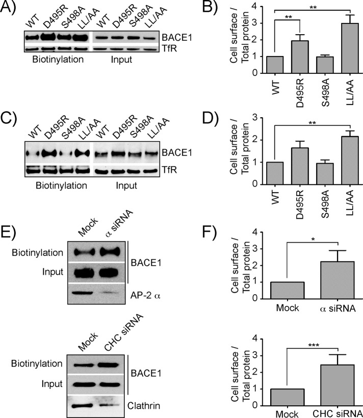
The β-site amyloid precursor protein (APP)-cleaving enzyme 1 (BACE1) is a transmembrane aspartyl protease that catalyzes the proteolytic processing of APP and other plasma membrane protein precursors. BACE1 cycles between the trans-Golgi network (TGN), the plasma membrane, and endosomes by virtue of signals contained within its cytosolic C-terminal domain. One of these signals is the DXXLL-motif sequence DISLL, which controls transport between the TGN and endosomes via interaction with GGA proteins. Here we show that the DISLL sequence is embedded within a longer [DE]XXXL[LI]-motif sequence, DDISLL, which mediates internalization from the plasma membrane by interaction with the clathrin-associated, heterotetrameric adaptor protein 2 (AP-2) complex. Mutation of this signal or knockdown of either AP-2 or clathrin decreases endosomal localization and increases plasma membrane localization of BACE1. Remarkably, internalization-defective BACE1 is able to cleave an APP mutant that itself cannot be delivered to endosomes. The drug brefeldin A reversibly prevents BACE1-catalyzed APP cleavage, ruling out that this reaction occurs in the endoplasmic reticulum (ER) or ER-Golgi intermediate compartment. Taken together, these observations support the notion that BACE1 is capable of cleaving APP in late compartments of the secretory pathway.


Figures

FIGURE 1:
FIGURE 1:
Analysis of the interaction of the BACE1 cytosolic tail with the AP-2 α-σ2 hemicomplex. (A) Schematic representation of BACE1, indicating its topological domains. CT, cytosolic tail; PP, propeptide; SP, signal peptide; TM, transmembrane domain. Also indicated are amino acid numbers, DTGS (residues 93–96), and DSGT (residues 289–292) that are necessary for β-secretase activity, and sequence of the C-terminal portion of the CT (residues 477–501), including overlapping DISLL (residues 496–500) and DDISLL signals (residues 495–500) that bind to GGA and AP-2, respectively. (B) Y3H interaction of the BACE1 cytosolic tail, HIV-1 Nef, and mouse tyrosinase (Tyr) tail with the AP-2 α-σ2 hemicomplex (Chaudhuri et al., 2007; Mattera et al., 2011). (C) Y3H analysis of the interaction of the BACE1 tail (left) and HIV-1 Nef (right) with the AP-1 γ-σ1A, AP-2 α-σ2, and AP-3 δ-σ3A hemicomplexes. (D) Y3H analysis of the interaction of BACE1 tail mutants with the AP-2 α-σ2 hemicomplex. The SV40 large T antigen (T-Ag) and p53 were used as controls. Growth in the absence of histidine (–His) is indicative of interactions.
FIGURE 2:
FIGURE 2:
Requirement of the [DE]XXXL[LI] signal for BACE1 localization to endosomes. (A) H4 human neuroglioma cells were transiently transfected with BACE1-wt and BACE1 mutant constructs for 12 h as indicated. Cells were fixed with 4% paraformaldehyde and processed as described (Burgos et al., 2010). Localization was determined by immunostaining using 1:100 dilution of monoclonal antibody (mAb) to HA, followed by incubation with a 1:1000 dilution of Alexa Fluor 594–labeled anti–mouse immunoglobulin G (IgG) antibody. Coverslips were mounted using Fluoromount-G and examined with an Olympus FluoView FV1000 laser scanning confocal unit attached to an Olympus IX81 motorized inverted microscope. (B) Rat hippocampal neurons were transfected with the same constructs on DIV-4. Cells were fixed with 4% paraformaldehyde and processed on DIV-7 as described in A. (C) Quantification of images from A was performed and scored as cytoplasmic puncta, plasma membrane, or intermediate phenotype (both plasma membrane and cytoplasmic puncta) and plotted as percentage of cells showing the respective phenotype. Values are the mean ± SD from six independent experiments. (D) Quantification of images from B was performed as in C. Values represent the mean ± SD from three independent experiments. ***p < 0.001. Bars, 10 μm.
FIGURE 3:
FIGURE 3:
Redistribution of BACE1 in H4 human neuroglioma cells upon siRNA-mediated knockdown of AP-2 or clathrin. Increased localization of BACE1 to the plasma membrane upon AP-2 α (A) or clathrin heavy chain (CHC) RNAi (C). Left, BACE1 localization, right, AP-2 α or CHC staining. BACE1 was immunostained with pAb to BACE1, whereas localization of clathrin and AP-2 were determined by staining with mAB to CHC (Thermo Scientific) and α-adaptin (Affinity Bioreagents), respectively. Bar, 10 μm. (B, D) Quantification of the phenotype carried out for images in A and C, respectively, by scoring for staining in various compartments, such as cytoplasmic puncta, plasma membrane, and intermediate (both plasma membrane and cytoplasmic puncta). Values are the mean ± SD from three experiments. ***p< 0.001.
FIGURE 4:
FIGURE 4:
Colocalization of BACE1 and TfR upon depletion of AP-2. Mock or AP-2 α siRNA-treated HeLa cells were transiently transfected with a plasmid encoding BACE1-wt. After 12 h of transfection, cells were incubated in culture medium deprived of serum for 1 h at 37°C to deplete the cells of endogenous transferrin. A subset of cells was the incubated either with 40 μg/ml Alexa Fluor 594–labeled human TfR or 1:100 dilution of mouse anti-HA antibody in culture medium without serum for 30 min at 4°C to stain cell surface BACE1-wt and transferrin receptor. Another subset of cells was incubated in culture medium without serum for 30 min at 37°C to allow labeled transferrin to reach a steady state. After treatment, cells were fixed with 4% paraformaldehyde and processed as in Figure 2A. Only cells treated at 37°C were further permeabilized and incubated with a 1:500 dilution of mouse anti-HA antibody for 1 h at 37°C. Both subsets of cells were incubated with a 1:1000 dilution of Alexa Fluor 488–labeled anti-mouse IgG antibody (Invitrogen) for 1 h at 37°C. Notice an almost complete colocalization between BACE1-wt and labeled transferrin at clathrin-coated pits at the cell surface in the presence of AP-2 (mock cells) and the disappearance of the punctate pattern in the AP-2 KD cells. Bar, 10 μm.
FIGURE 5:
FIGURE 5:
Cell surface expression and internalization of wild-type and mutant BACE1 constructs. (A) H4 human neuroglioma cells transiently transfected either with BACE1-wt or BACE1 mutant constructs were surface labeled with Sulfo-NHS-LC-Biotin at 4°C. After lysis, the extracts were incubated with NeutrAvidin-agarose, and bound proteins were analyzed by SDS–PAGE and immunoblotting using antibodies to an extracellular HA epitope tag and the TfR. (B) Densitometric quantification of bands was performed on three independent experiments as shown in A. Values are the mean ± SD from three independent experiments after normalization to wt in each experiment. (C) Rat cortical neurons were transfected with wild-type and mutant BACE1 constructs on DIV-4 and subjected to surface labeling by biotinylation and isolation of biotinylated proteins on DIV-7. Analysis was performed as described in A. (D) Quantification for three independent experiments as in B. (E) H4 human neuroglioma cells transiently transfected with wild-type BACE1 construct were subjected to KD of either the large subunit of AP-2 (α siRNA) or clathrin heavy chain (CHC siRNA). Bottom, KD efficiency, using specific antibodies to the α-adaptin subunit for AP-2 complex and CHC for clathrin. Cell surface-biotinylation against total protein was calculated and processed as described in A. (F) Quantification for three independent experiments as in B. *p < 0.05, **p < 0.01, ***p < 0.001.
FIGURE 6:
FIGURE 6:
Internalization of wild-type and mutant BACE1. (A) H4 human neuroglioma cells transiently transfected with BACE1-wt or mutant constructs were surface labeled with Sulfo-NHS-LC-Biotin at 4°C and chased for 0, 30, and 60 min at 37°C in complete medium. After the chase period, the remaining surface label was specifically cleaved with non–cell-permeable glutathione. Isolation of biotinylated protein and analysis was performed as described in the legend to Figure 4. (B) H4 cells transiently transfected with wild-type and mutant BACE1 constructs were surface labeled at 4°C with polyclonal antibodies to the extracellular HA epitope tag, washed, and then chased for 20 min in complete medium before fixation. Internalization was analyzed by indirect immunofluorescence using Alexa 594–conjugated secondary antibody. (C) Images from B were quantified for localization of internalized BACE1 to cytoplasmic puncta (endosomal), plasma membrane, or intermediate phenotype (both plasma membrane and cytoplasmic puncta) and plotted as percentage of cells. Values are the mean ± SD from three different experiments. ***p < 0.001. (D) Antibody uptake assay carried out upon knockdown on AP-2 α and CHC as described in B. Bars, 10 μm.
FIGURE 7:
FIGURE 7:
BACE1-catalyzed APP cleavage is independent of BACE1 localization to endosomes. (A) Schematic representation of APP-EFGP, indicating the position of the Aβ peptide, N- and C-terminal fragments (NTF and CTF, respectively), transmembrane domain (TM), the α-, β-, β′- and γ-secretase, caspase-cleavage sites and the resulting fragments, and mutations or inhibitors that block cleavage. Underlined is the region in APP recognized by the 6E10 antibody used in this study to identify C99; all other CTFs and APP-FL were detected using anti–GFP-HRP antibody. The γ-secretase inhibitor DAPT was added to the media posttransfection for experiments in B–F. (B) Cleavage of an APP construct with mutations in the α-secretase and caspase-cleavage sites (APP-F/P-D/A) was analyzed in cells cotransfected with constructs encoding wild-type BACE1, an inactive form of BACE1 (D289N), and wild-type BACE1 in the presence of β-secretase inhibitor (β-INH) or without BACE1 (–). Cell extracts were analyzed by SDS–PAGE and immunoblotting for α-, β-, and β′-secretase cleavage products. C89 was the prominent band detected upon expression of wild-type BACE1 but not under the other conditions. (C) H4 cells were treated with siRNA to AP-2 α or mock treated, and 72 h after RNAi cells were transfected with a wild-type BACE1 construct. Analysis was performed as in B. (D) H4 human neuroglioma cells were transiently cotransfected with APP-F/P-D/A and with BACE1-wt or endocytosis-defective BACE1 mutants or in the presence of BACE1 mutant deficient in β-cleavage constructs, as indicated. A control was carried out in parallel in the absence of BACE1 overexpression as shown. Analysis was performed as in B, using antibodies to detect APP-FL and C89 fragment as mentioned, and BACE1 was detected using anti–HA-HRP antibody. (E) Densitometric quantification of bands was performed on three independent experiments such as that shown in D. The mean ± SD of the ratio of C89 fragment to full-length APP was calculated. (F) Rat cortical neurons were cotransfected with APP-F/P-D/A and BACE1-wt and BACE1 mutant constructs at DIV-4. Controls involving coexpression of APP-F/P-D/A with inactive BACE1 D289N mutant or wild-type BACE1 in the presence of β-secretase inhibitor were included in the experiment. Analysis was performed on DIV-7 as described in D. (G) Quantification from three independent experiments was performed as in E and represented as mean ± SD.
FIGURE 8:
FIGURE 8:
APP is cleaved by BACE1 independent of endosomal localization. (A) H4 human neuroglioma cells were transfected with constructs encoding APP-F/P-D/A or APP-F/P-D/A with additional mutation of all three tyrosine residues in the cytosolic tail (APP 3Y-F/P-D/A), both expressing C-terminal GFP. Cells were stained for TGN46 and examined by fluorescence microscopy. Merging red and green channels generated the third picture on each row; yellow indicates overlapping localization. Insets show 3× magnifications. (B) Immunofluorescence images from A were quantified for Golgi localization. Values are the mean ± SD from three different experiments. ***p < 0.001. (C) Cells were biotinylated on the cell surface as described in Figure 5A and proteins analyzed by immunoblotting with an anti–GFP-HRP antibody. (D) Rat cortical neurons were cotransfected with APP 3Y-F/P-D/A and wild-type or mutant BACE1 constructs on DIV-4 and analyzed on DIV-7 as described in Figure 7E. Bar, 10 μm.
FIGURE 9:
FIGURE 9:
Cleavage of APP by BACE1 occurs in post-ER compartments. (A) H4 cells were transiently cotransfected with constructs encoding APP-F/P-D/A and BACE1-wt or mutants. Cells were incubated with DAPT in the presence or absence of BFA for 10 h posttransfection as indicated. Total extracts were subjected to SDS–PAGE and analyzed by immunoblotting with specific antibodies. Notice the generation of the C89 fragment by BACE1-wt and BACE1 mutants in the absence of BFA but not in the presence of BFA. (B) H4 cells were cotransfected with APP-F/P-D/A and BACE1-wt construct and maintained for 10 h in the presence of DAPT plus or minus BFA as indicated. BFA was washed out, and cells were further incubated for 0, 5, and 10 h in the presence of DAPT. Total extracts were analyzed by SDS–PAGE and immunoblotting as in A.

Similar articles

Cited by

References

    1. Bonifacino JS. The GGA proteins: adaptors on the move. Nat Rev Mol Cell Biol. 2004;5:23–32. - PubMed
    1. Bonifacino JS, Traub LM. Signals for sorting of transmembrane proteins to endosomes and lysosomes. Annu Rev Biochem. 2003;72:395–447. - PubMed
    1. Boucrot E, Saffarian S, Zhang R, Kirchhausen T. Roles of AP-2 in clathrin-mediated endocytosis. PLoS One. 2010;5:e10597. - PMC - PubMed
    1. Burgos PV, Mardones GA, Rojas AL, daSilva LL, Prabhu Y, Hurley JH, Bonifacino JS. Sorting of the Alzheimer's disease amyloid precursor protein mediated by the AP-4 complex. Dev Cell. 2010;18:425–436. - PMC - PubMed
    1. Chaudhuri R, Lindwasser OW, Smith WJ, Hurley JH, Bonifacino JS. Downregulation of CD4 by human immunodeficiency virus type 1 Nef is dependent on clathrin and involves direct interaction of Nef with the AP2 clathrin adaptor. J Virol. 2007;81:3877–3890. - PMC - PubMed

Publication types

MeSH terms