. 2012 Jul;23(13):2527-36.
doi: 10.1091/mbc.E12-01-0077. Epub 2012 May 2.

Local control of phosphatidylinositol 4-phosphate signaling in the Golgi apparatus by Vps74 and Sac1 phosphoinositide phosphatase

Affiliations

Local control of phosphatidylinositol 4-phosphate signaling in the Golgi apparatus by Vps74 and Sac1 phosphoinositide phosphatase

Christopher S Wood et al. Mol Biol Cell. 2012 Jul.

Abstract

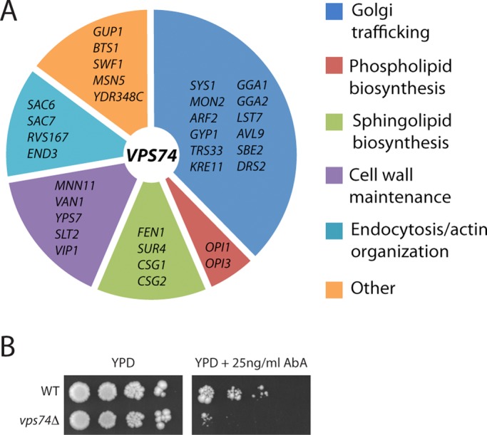
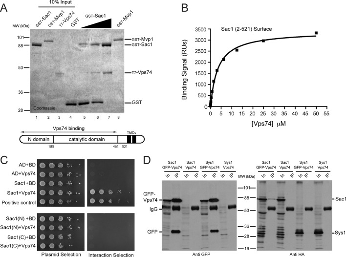
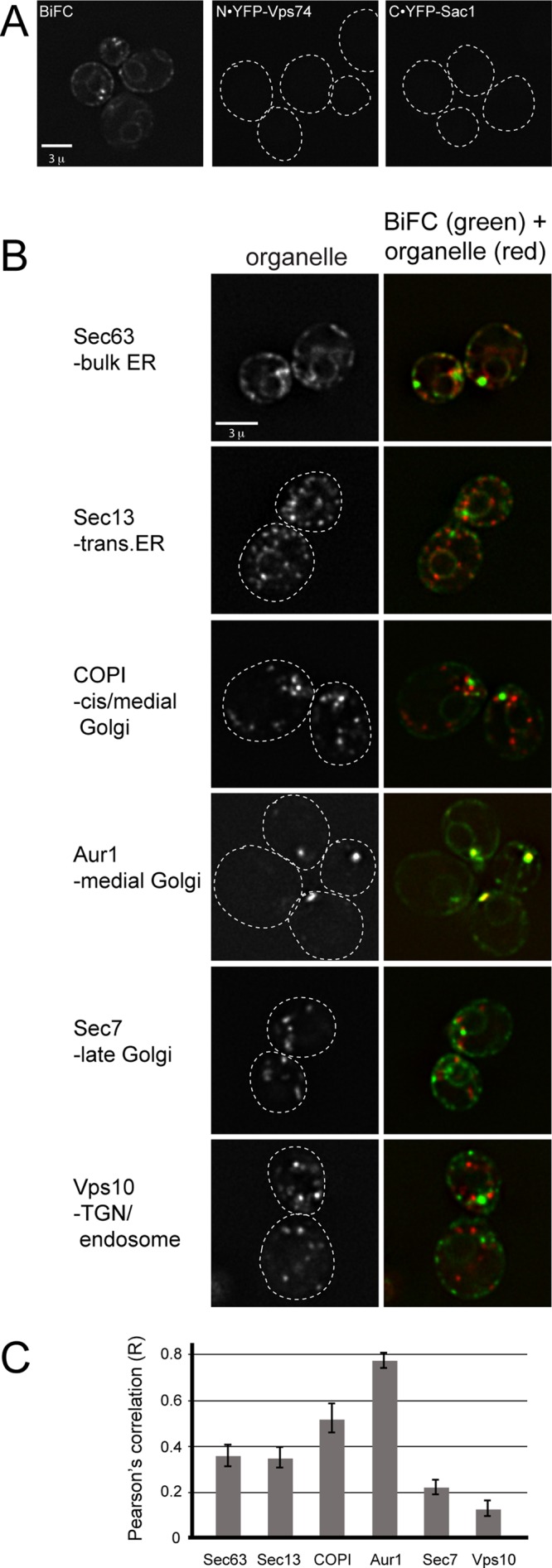
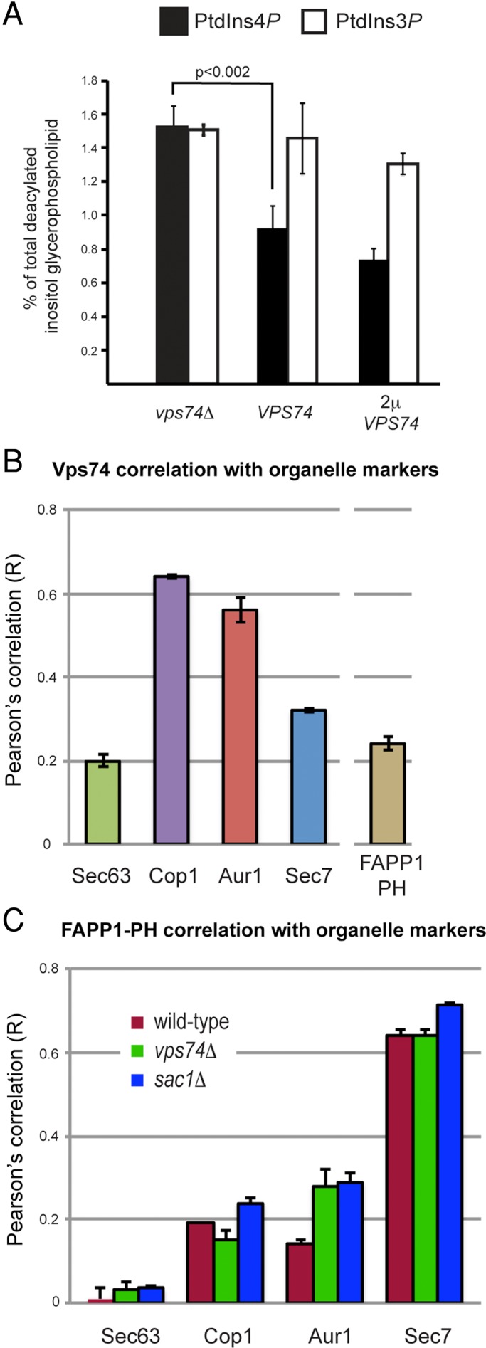
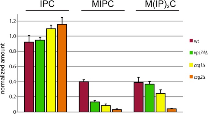
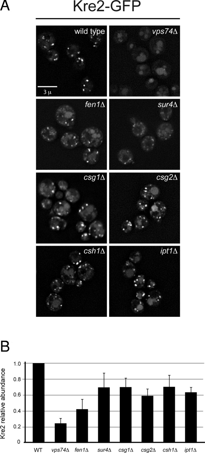
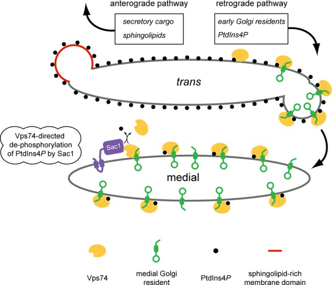
In the Golgi apparatus, lipid homeostasis pathways are coordinated with the biogenesis of cargo transport vesicles by phosphatidylinositol 4-kinases (PI4Ks) that produce phosphatidylinositol 4-phosphate (PtdIns4P), a signaling molecule that is recognized by downstream effector proteins. Quantitative analysis of the intra-Golgi distribution of a PtdIns4P reporter protein confirms that PtdIns4P is enriched on the trans-Golgi cisterna, but surprisingly, Vps74 (the orthologue of human GOLPH3), a PI4K effector required to maintain residence of a subset of Golgi proteins, is distributed with the opposite polarity, being most abundant on cis and medial cisternae. Vps74 binds directly to the catalytic domain of Sac1 (K(D) = 3.8 μM), the major PtdIns4P phosphatase in the cell, and PtdIns4P is elevated on medial Golgi cisternae in cells lacking Vps74 or Sac1, suggesting that Vps74 is a sensor of PtdIns4P level on medial Golgi cisternae that directs Sac1-mediated dephosphosphorylation of this pool of PtdIns4P. Consistent with the established role of Sac1 in the regulation of sphingolipid biosynthesis, complex sphingolipid homeostasis is perturbed in vps74Δ cells. Mutant cells lacking complex sphingolipid biosynthetic enzymes fail to properly maintain residence of a medial Golgi enzyme, and cells lacking Vps74 depend critically on complex sphingolipid biosynthesis for growth. The results establish additive roles of Vps74-mediated and sphingolipid-dependent sorting of Golgi residents.


Figures

FIGURE 1:
FIGURE 1:
Genetic profiling of vps74Δ cells. (A) Gene deletions that aggravate cell growth when combined with the vps74Δ allele are listed and categorized by their functions. (B) vps74Δ cells are hypersensitive to aureobasidin A, an inhibitor of complex sphingolipid biosynthesis. Tenfold serial dilutions of wild-type and vps74Δ cells were spotted onto YPD medium containing 25 ng/ml aureobasidin A and grown for 4 d.
FIGURE 2:
FIGURE 2:
Vps74 and Sac1 form a complex. (A) Purified Vps74 binds immobilized GST-Sac1 phosphatase domain. A series of binding reactions was set up in which the amount of immobilized GST-Sac1 (amino acids 2–521, comprising the entire N-terminal cytoplasmic portion) fusion protein was increased (lanes 5–7) while keeping the total amount of immobilized bait protein constant by adding a compensatory amount of GST. The position of T7- and His-tagged Vps74, confirmed by anti-T7 immunoblotting (unpublished data), is indicated. Note that Vps74 does not bind to GST-Mvp1, an endosomal protein that serves as a specificity control. A schematic of Sac1 structure is shown below the gel, with the amino acid positions of the domains indicated. (B) SPR analysis of Vps74 binding to Sac1 (amino acids 2–251). A series of samples of Vps74 at the indicated concentrations was passed over a CM5 chip to which Sac1 had been immobilized. The curve indicates the fit of a representative data set to a simple one-site binding equation. The KD value, determined from the fit to at least three independent data sets, is 3.8 ± 0.4 μM. (C) Yeast two-hybrid analysis of Vps74–Sac1 interaction. Cells containing plasmids that express the indicated proteins were spotted by limited dilution (1:10) onto nonselective plates (left) and onto interaction-selective plates (right) containing 10 mM 3-amino-triazole. The plates were photographed after 2 d incubation at 30°C. All Sac1-derived constructs were expressed as C-terminal fusions to the Gal4 activation domain. “Sac1” refers to amino acids 1–461 (encompassing the entire Sac1 homology region), “Sac1(N)” indicates the N-terminal subdomain (amino acids 1–185), and “Sac1(C)” indicates the catalytic subdomain (amino acids 186–421) of the Sac1 homology region. A schematic diagram of Sac1 summarizes the positions of the structural features of Sac1. The positive-control assay consisted of a fragment of Pan1 (amino acids 96–715) and Yap180 (amino acids 432–637) (Wendland and Emr, 1998). (D) Coimmunopurification of GFP-Vps74 and Sac1-HA. Cells expressing GFP-Vps74 and HA epitope-tagged Sac1 (Sac1-HA), or Sys1-HA as a control, were converted to spheroplasts and incubated with a bifunctional, cleavable cross-linking reagent. After the cells were detergent-solubilized, GFP-Vps74 was immunopurified with anti-GFP antiserum, and the bound material was probed with anti-HA antiserum. In the lanes marked “In,” 1% of the material used for the immunopurification is loaded. The positions of molecular-size standards (kilodaltons) are indicated.
FIGURE 3:
FIGURE 3:
Visualization of Vps74-Sac1 complexes in vivo. (A) Identification of Vps74-Sac1 complexes in vivo by BiFC. Fluorescence micrographs of cells expressing the N-terminal portion of YFP fused to the N-terminus of Vps74 (N·YFP-Vps74), and the C-terminal portion of YFP fused to the N-terminus of Sac1 (C·YFP-Sac1), or both fusion proteins (BiFC) are shown. Fluorescence complementation is observed when both proteins are coexpressed. (B) Vps74-Sac1 BiFC colocalization with secretory organelles. The indicated resident proteins of the ER and Golgi were tagged on their C-termini with mKate in cells expressing N·YFP-Vps74and C·YFP-Sac1 and visualized by deconvolution microscopy. One optical focal plane from one Z-stack is shown as an example. (C) Pearson's correlation coefficients (r) were determined for the entire volumes (i.e., voxels) of at least 25 cells in at least three different fields. The mean r values (± SE) are plotted.
FIGURE 4:
FIGURE 4:
PtdIns4P is elevated in the Golgi of vps74Δ cells. (A) Elevated PtdIns4P in vps74Δ cells. Cells were labeled to steady state with [3H]myoinositol and the amounts of PtdIns4P and PtdIns3P (as a reference) were determined by high-performance liquid chromatography analysis of the corresponding deacylated glycero-phosphoinositol-phosphate derivatives. The analysis was conducted in a sec14-1 kes1Δ mutant background with the indicated VPS74 genotype to facilitate accurate determinations of changes in PtdIns4P levels. (B) Vps74 is enriched on cis and medial compartments of the Golgi. The distribution of GFP-Vps74 across ER and Golgi compartments was determined by calculating Pearson's correlation coefficients (r) for GFP-Vps74 and the indicated ER and Golgi residents expressed with a C-terminal fusion to mKate. A similar analysis was carried to compare colocalization of mCherry-Vps74 and GFP-FAPP1·PH. The mean r values (± SE) are plotted. (C) Distribution of the GFP-FAPP1·PH PtdIns4P probe across ER and Golgi compartments. GFP-FAPP1·PH was expressed from a low-copy CEN plasmid in strains expressing the mKate-tagged Sec63 (ER), Cop1, Aur1, or Sec7, and Pearson's correlation coefficients (r) were determined for voxels from deconcolved Z-series micrographs. The mean r values (± SE) are plotted.
FIGURE 5:
FIGURE 5:
Complex sphingolipid analyses. The amount of each of the indicated sphingolipid species (normalized to phosphatidylinositol) in the indicated strains is plotted. The height of each bar represents the mean of three independent determinations.
FIGURE 6:
FIGURE 6:
Disrupted localization of the medial Golgi mannosyltransferase, Kre2, in sphingolipid mutants. (A) Kre2-GFP was visualized in the indicated sphingolipid pathway mutants by deconvolution microscopy. One representative image from the Z-series is shown. For comparison, wild-type and the vps74Δ cells are shown. (B) Steady-state abundance of myc epitope–tagged Kre2 (Kre2-myc) in the indicated strains was determined by immunoblotting of detergent cell lysates. The means of three independent measurements are plotted, normalized to wild-type cells, with the SE of the measurements indicated.
FIGURE 7:
FIGURE 7:
Model of Vps74-mediated protein sorting and PtdIns4P signaling in the Golgi apparatus. The medial and trans compartments of the Golgi apparatus are depicted with PtdIns4P (black circles) most abundant on the trans cisterna. Vps74 (yellow oval) recognizes PtdIns4P and early Golgi-resident mannosyltransferases (green). Vps74 is depicted to sort these enzymes into COPI-coated retrograde vesicles (COPI coat is not depicted). Also in the trans cisterna, complex sphingolipids are preferentially packaged with secretory cargo into anterograde-directed transport vesicles (red vesicle). We propose that the efficiency of Vps74-mediated sorting of Golgi residents into the retrograde pathway increases as a consequence of excluding Golgi residents from sphingolipid-rich domains. As a consequence of copackaging PtdIns4P with Golgi residents into retrograde vesicles, PtdIns4P is delivered to the medial cisterna. Sac1 (purple) cycles between the ER and Golgi apparatus and the data presented herein indicate that Sac1 and Vps74 function in the Golgi to maintain a low amount of PtdIns4P on the medial cisterna; we propose that this is mediated by the Vps74-Sac1 complex.

Similar articles

Cited by

References

    1. Breslow DK, Collins SR, Bodenmiller B, Aebersold R, Simons K, Shevchenko A, Ejsing CS, Weissman JS. Orm family proteins mediate sphingolipid homeostasis. Nature. 2010;463:1048–1053. - PMC - PubMed
    1. Bretscher MS, Munro S. Cholesterol and the Golgi apparatus. Science. 1993;261:1280–1281. - PubMed
    1. Brice SE, Alford CW, Cowart LA. Modulation of sphingolipid metabolism by the phosphatidylinositol-4-phosphate phosphatase Sac1p through regulation of phosphatidylinositol in Saccharomyces cerevisiae. J Biol Chem. 2009;284:7588–7596. - PMC - PubMed
    1. Cheong FY, Sharma V, Blagoveshchenskaya A, Oorschot VM, Brankatschk B, Klumperman J, Freeze HH, Mayinger P. Spatial regulation of Golgi phosphatidylinositol-4-phosphate is required for enzyme localization and glycosylation fidelity. Traffic. 2010;11:1180–1190. - PMC - PubMed
    1. Cleves AE, Novick PJ, Bankaitis VA. Mutations in the SAC1 gene suppress defects in yeast Golgi and yeast actin function. J Cell Biol. 1989;109:2939–2950. - PMC - PubMed

Publication types

MeSH terms

Substances