. 2012 Jul;11(7):1400-10.
doi: 10.1158/1535-7163.MCT-12-0172. Epub 2012 May 2.

Human monoclonal antibodies targeting nonoverlapping epitopes on insulin-like growth factor II as a novel type of candidate cancer therapeutics

Affiliations

Human monoclonal antibodies targeting nonoverlapping epitopes on insulin-like growth factor II as a novel type of candidate cancer therapeutics

Weizao Chen et al. Mol Cancer Ther. 2012 Jul.

Abstract

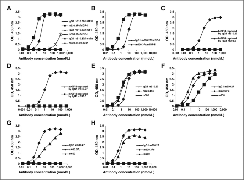
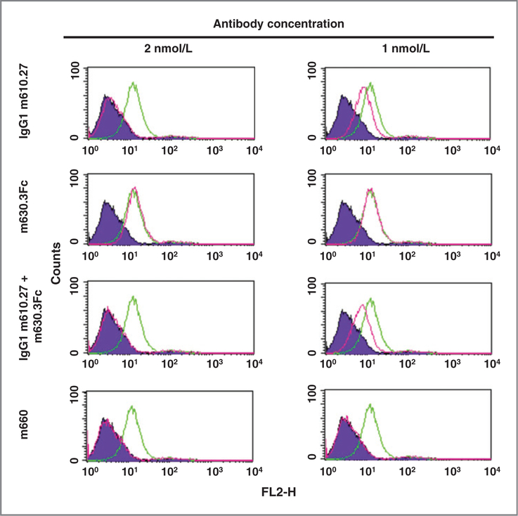
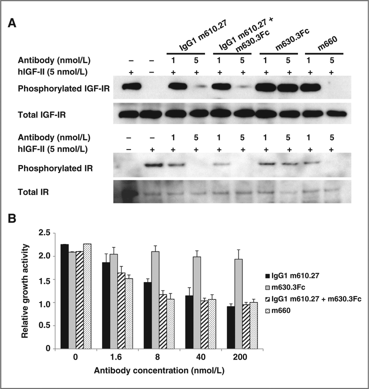
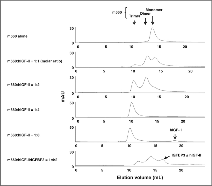
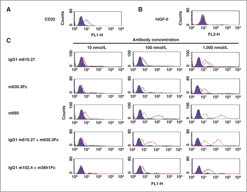
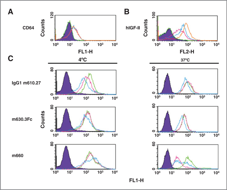
Soluble ligands are important targets for therapy of cancers and other diseases. Therapeutic monoclonal antibodies (mAb) against such ligands block their interactions with corresponding receptors but do not enhance their removal from the circulation and can increase their half-lives because of the long half-lives of the antibodies. We have hypothesized that mAbs targeting two or more nonoverlapping epitopes on the same ligand could form oligomeric antibody-ligand complexes that can bind to cells expressing Fc gamma receptors (FcγRs) with high avidity leading to their fast and irreversible removal from the circulation. Insulin-like growth factor II (IGF-II) is an example of such ligands and an important target for human cancer therapy. We identified two mAbs, m610.27 and m630.3, which bound to nonoverlapping epitopes on IGF-II with nanomolar affinity, and generated a bispecific antibody, m660. m660 inhibited the interaction of human IGF-II (hIGF-II) with the human breast cancer cell line MCF-7, hIGF-II-mediated IGF receptor type I and insulin receptor phosphorylation, and cell growth. In the presence of hIGF-II, large complexes of m660 were formed that bound to FcγRII-expressing BJAB cells much more efficiently than the monospecific antibody-hIGF-II complexes and were presumably phagocytosed by phorbol 12-myristate 13-acetate-stimulated macrophage-like U937 cells. A mixture of m610.27 and m630.3 exhibited similar properties. To our knowledge, these mAbs are the first reported to target nonoverlapping epitopes on a cancer-related ligand and could represent a novel class of candidate therapeutics against cancers. This approach could also be used to irreversibly eliminate other disease-related soluble ligands.


Conflict of interest statement

Disclosure of Potential Conflicts of Interest

No potential conflicts of interest were disclosed.

Figures

Figure 1.
Figure 1.
Binding and competition ELISA. A, binding of IgG1 m610.27 and m630.3Fc to hIGF-II, hIGF-I, and human insulin. B, binding of the antibodies to mIGF-II. C, competition of m630.3Fc with IgG1s m610.27 and m708.5 in binding to hIGF-II. D, competition of m630.3Fc with IgG1s m610.27 and m708.5 in binding to mIGF-II. E, binding of m660 to hIGF-II. F, binding of m660 to long hIGF-II. G, competition of the antibodies with IGFBP2 in binding to hIGF-II. H, competition of the antibodies with IGFBP3 in binding to hIGF-II. All assays were done in duplicate. Error bars were included in all binding curves.
Figure 2.
Figure 2.
Inhibition of FACS binding of hIGF-II to MCF-7 cells. Diagrams for reference cells, which were incubated with streptavidin-PE conjugate only, are in purple. Diagrams for cells incubated with hIGF-II only are in green. Those for hIGF-II plus antibodies are in pink.
Figure 3.
Figure 3.
Inhibition of hIGF-II–mediated IGF-IR and IR phosphorylation (A), and MCF-7 cell growth (B). In the phosphorylation assay, MCF-7 cells were starved in serum-free medium for 5 hours, followed by addition of treatment medium containing 5 nmol/L hIGF-II without or with the antibodies at different concentrations. After 30-minute incubation, cells were chilled and lysed. IGF-IR or IR was immunoprecipitated, and the phosphorylated receptor was detected with a phospho-tyrosine–specific antibody. The membranes were stripped and reprobed by the same polyclonal antibody used for the immunoprecipitation to detect the total amount of the receptors. In the cell growth assay, mean relative light units (RLU) for duplicate wells were determined. Relative growth activity of the cells was calculated by the following formula: average RLU of hIGF-II–containing wells/average RLU of hIGF-II–free wells.
Figure 4.
Figure 4.
Size exclusion chromatography analysis of m660–hIGF-II complexes. The bold arrows shown at the top indicate the positions where a monomer, dimer, or trimer of m660 should elute. The bold arrows at the bottom indicate the peaks corresponding to the elution of free hIGF-II and IGFBP3 alone or in complex with hIGF-II, respectively. The ± denotes with or without.
Figure 5.
Figure 5.
FACS binding of antibody–hIGF-II complexes to BJAB cells. A, expression of FcγRII on BJAB cells. The diagram for reference cells is in purple. The diagram for cells incubated with 1:100 (v/v) diluted FITC-conjugated mouse anti-human CD16 (FcγRIII) antibody is in green. The pink diagram is for cells incubated with the anti-human CD32 (FcγRII) antibody at the same dilution. B, binding of biotinylated hIGF-II to BJAB cells. The green diagram indicates the cells incubated with streptavidin-PE conjugate only. The diagrams for cells incubated with hIGF-II at concentrations of 10, 100, and 1,000 nmol/L are in pink, blue, and orange, respectively. C, interactions of antibody–hIGF-II complexes with BJAB cells. The diagrams for reference cells are in purple. The green diagrams are for cells incubated with antibody alone. The pink diagrams are for cells incubated with a mixture of antibody and hIGF-II at a molar ratio of 1:2 (for monospecific antibodies) or 1:4 (for m660).
Figure 6.
Figure 6.
FACS binding of m660–hIGF-II complexes to PMA-stimulated U937 cells. A, expression of FcγRI on U937 cells. The diagram for reference cells that were not stimulated is in purple. The diagram for cells mock-stimulated with PMA solvent dimethyl sulfoxide (DMSO) is in green. The pink, blue, and orange diagrams are for cells stimulated with 10, 20, and 30 ng/mL PMA, respectively. B, binding of biotinylated hIGF-II to U937 cells. The green diagram indicates cells incubated with streptavidin-PE conjugate only. The diagrams for cells incubated with hIGF-II at concentrations of 10, 100, and 1,000 nmol/L are in pink, blue, and orange, respectively. C, interactions of antibody–hIGF-II complexes with U937 cells. The purple diagram is for reference cells incubated with the secondary antibody only. The diagrams for cells incubated with 10 nmol/L antibody alone, 10 nmol/L antibody plus 20 (for monospecific antibodies), or 40 nmol/L (for m660) hIGF-II, and an combination of the antibody, hIGF-II, and 50 μmol/L cytochalasin D are in green, pink, and blue, respectively.

References

    1. Ryan PD, Goss PE. The emerging role of the insulin-like growth factor pathway as a therapeutic target in cancer. Oncologist 2008;13:16–24. - PubMed
    1. Samani AA, Yakar S, LeRoith D, Brodt P. The role of the IGF system in cancer growth and metastasis: overview and recent insights. Endocr Rev 2007;28:20–47. - PubMed
    1. Hendrickson AW, Haluska P. Resistance pathways relevant to insulin-like growth factor-1 receptor-targeted therapy. Curr Opin Investig Drugs 2009;10:1032–40. - PubMed
    1. Goya M, Miyamoto S, Nagai K, Ohki Y, Nakamura K, Shitara K, et al. Growth inhibition of human prostate cancer cells in human adult bone implanted into nonobese diabetic/severe combined immunodeficient mice by a ligand-specific antibody to human insulin-like growth factors. Cancer Res 2004;64:6252–8. - PubMed
    1. Feng Y, Zhu Z, Xiao X, Choudhry V, Barrett JC, Dimitrov DS. Novel human monoclonal antibodies to insulin-like growth factor (IGF)-II that potently inhibit the IGF receptor type I signal transduction function. Mol Cancer Ther 2006;5:114–20. - PubMed

Publication types

MeSH terms