. 2012 Jul;40(Web Server issue):W222-7.
doi: 10.1093/nar/gks363. Epub 2012 May 2.

MOLEonline 2.0: interactive web-based analysis of biomacromolecular channels

Affiliations

MOLEonline 2.0: interactive web-based analysis of biomacromolecular channels

Karel Berka et al. Nucleic Acids Res. 2012 Jul.

Abstract

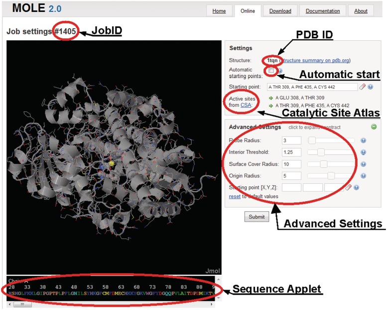
Biomolecular channels play important roles in many biological systems, e.g. enzymes, ribosomes and ion channels. This article introduces a web-based interactive MOLEonline 2.0 application for the analysis of access/egress paths to interior molecular voids. MOLEonline 2.0 enables platform-independent, easy-to-use and interactive analyses of (bio)macromolecular channels, tunnels and pores. Results are presented in a clear manner, making their interpretation easy. For each channel, MOLEonline displays a 3D graphical representation of the channel, its profile accompanied by a list of lining residues and also its basic physicochemical properties. The users can tune advanced parameters when performing a channel search to direct the search according to their needs. The MOLEonline 2.0 application is freely available via the Internet at http://ncbr.muni.cz/mole or http://mole.upol.cz.


Figures

Figure 1.
Figure 1.
MOLEonline 2.0 setup webpage for channel calculation. Each job is assigned a job ID to allow easy access to the results. Setup starts with the selection of a PDB file (here 1TQN) either from the PDB database or uploaded as a user file. The tunnel starting point can be selected automatically (inside cavities detected by MOLE 2.0 algorithm) or manually, by using CSA (37), via selection through the interactive sequence applet on the bottom of the page or by specifying of x, y, z coordinates in advanced settings. Advanced settings also enable the adjustment of parameters determining the tunnel searching algorithm. All parameters are set in Ångströms (for details see the text).
Figure 2.
Figure 2.
Results of channel analysis of Cytochrome P450 3A4 (CYP3A4) using the setup shown in Figure 1. Four channels found from user-specified starting point are shown, whereas the automatic detection also found additional 17 tunnels which are not shown for clarity. The profile of the tunnel #1 along the centerline and list of lining residues are shown in the external windows (right-hand side). A list of all the unique lining residues and the corresponding side chains alone is displayed along with physicochemical properties of the respective channel. Lining residues can also be visualized along the channel centerline, with the channel represented by maximally inscribed spheres in the Jmol window. It is also possible to show molecular surface and all detected cavities and their volumes. In addition, starting points can be shown as small cubes for original user-defined starting point (in magenta), for optimized position of such starting point (in green) and for all automatically detected starting points (in yellow). Information about tunnel profiles and lining residues can be further exported in form of report, PDB file or python file for visualization in Pymol.
Figure 3.
Figure 3.
Visualization of ribosomal exit tunnel (RET) of a large ribosomal unit from Haloarcula marismortui (PDB: 1JJ2). The figure was prepared in Pymol using an exported python file containing positions of all the channels identified by MOLEonline. Only the RET is shown and ribosomal proteins L4 (green), L22 (blue), L39E (red) lining the tunnel are highlighted. The channel profile shows the positions of three bottlenecks.

References

    1. Walz T, Smith BL, Agre P, Engel A. The 3-dimensional structure of human erythrocyte aquaporin chip. EMBO J. 1994;13:2985–2993. - PMC - PubMed
    1. Engel A, Fijiyoshi Y, Agre P. The importance of aquaporin water channel protein structures. EMBO J. 2000;19:800–806. - PMC - PubMed
    1. Jiang YX, Lee A, Chen JY, Cadene M, Chait BT, MacKinnon R. Crystal structure and mechanism of a calcium-gated potassium channel. Nature. 2002;417:515–522. - PubMed
    1. Doyle DA, Cabral JM, Pfuetzner RA, Kuo AL, Gulbis JM, Cohen SL, Chait BT, MacKinnon R. The structure of the potassium channel: molecular basis of K+ conduction and selectivity. Science. 1998;280:69–77. - PubMed
    1. Gouaux E, MacKinnon R. Principles of selective ion transport in channels and pumps. Science. 2005;310:1461–1465. - PubMed

Publication types