. 2012 Jun 1;4(6):a009654.
doi: 10.1101/cshperspect.a009654.

Signaling pathways in cell polarity

Affiliations

Signaling pathways in cell polarity

Luke Martin McCaffrey et al. Cold Spring Harb Perspect Biol. .

Abstract

A key function of signal transduction during cell polarization is the creation of spatially segregated regions of the cell cortex that possess different lipid and protein compositions and have distinct functions. Polarity can be initiated spontaneously or in response to signaling inputs from adjacent cells or soluble factors and is stabilized by positive-feedback loops. A conserved group of proteins, the Par proteins, plays a central role in polarity establishment and maintenance in many contexts. These proteins generate and maintain their distinct locations in cells by actively excluding one another from specific regions of the plasma membrane. The Par signaling pathway intersects with multiple other pathways that control cell growth, death, and organization.


Figures

Figure 1.
Figure 1.
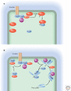
(A) Extrinsic signals normally are responsible for driving cell polarization, although it can also occur spontaneously under certain conditions. (B) Cell polarization is established by signal transduction pathways that spatially segregate different regions of the cell, especially the cell cortex, and this organization is reinforced and maintained by positive-feedback loops.
Figure 2.
Figure 2.
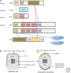
(A) Schematic showing domain structures of Par polarity proteins and their interactions. Phox and Bem1 domain (PB1), forms homodimers and heterodimers; zinc finger domain (Zn); PSD95, Dlg1, ZO-1 domain (PDZ), binds other PDZ domains and carboxy-terminal peptide motifs; conserved region domain (CR1), forms homo-oligomers; atypical protein kinase C binding domain (aPKCBD); ubiquitin-binding-associated domain (UBA); kinase-associated domain (KA). (B) The different distributions of these polarity proteins in an epithelial cell and a neuroblast stem cell, together with the localization of other interacting proteins. Note that whereas in neuroblasts all of the polarity proteins form a complex (the “Par complex”) at the apical cortex, this is not the case in epithelial cells, in which Par3 is not associated with Par6 and aPKC but is associated instead with the tight junction complex. The orientation of the mitotic spindle is controlled by the Par proteins and is different in neuroblasts (vertical) versus epithelial cells (horizontal). This difference reflects the distinct functions of polarity in the two cell types: segregation of cell fate determinants into only one daughter cell in the neuroblast versus formation of a polarized sheet of cells by the epithelium.
Figure 3.
Figure 3.
Positive-feedback loops that drive polarization of budding yeast during cell division. Cdc42 can cycle between GDP- and GTP-bound states, catalyzed by guanine nucleotide exchange factors (GEFs) that load GTP onto the protein, and GTPase-activating proteins (GAPs) that stimulate hydrolysis of the bound GTP. In the actin/Cdc42 loop, local enrichment of Cdc42-GTP at the cell cortex triggers nucleation of actin cables, along which vesicles recruit more Cdc42, which nucleates more actin cables. In the Cdc42/adaptor/GEF loop, local enrichment of Cdc42-GTP recruits an adaptor protein (Bem1), which, in turn, recruits a GEF for Cdc42 (Cdc24), which produces more Cdc42-GTP, which can, in turn, recruit more Bem1 and Cdc24.
Figure 4.
Figure 4.
Mechanisms for the transport, cortical association, and anchoring of Par3 in mammalian cells. It is not yet known if all of these mechanisms operate in any one cell type, and additional processes, such as RNA localization, might play roles in certain circumstances. Junctional adhesion molecule (JAM) is shown as an example of a transmembrane protein to which Par3 can be anchored, but others exist, such as the neurotrophin receptor, p75NTR, in mammalian Schwann cells (Chan et al. 2006). PP1α is a phosphatase.
Figure 5.
Figure 5.
Active exclusion from cortical domains. A common mechanism for the establishment of discrete cortical regions of the cell occurs through the active removal of unwanted proteins from within a particular region. A kinase within the region phosphorylates the protein, resulting in the association of Par5 (14-3-3), which displaces the protein from the cell cortex. (A) Par1 is restricted to the lateral membranes in epithelial cells. Any Par1 that strays onto the apical surface is phosphorylated by aPKC, which recruits Par5 (14-3-3), causing its disassociation from the membrane. (B) aPKC at the apical surface of an epithelial cell phosphorylates any Pins (also known as LGN) protein that binds to Gαi within this domain. Association of the phosphorylated Pins with 14-3-3 triggers its dissociation from Gαi. Because phosphorylation does not occur at the basolateral membrane, Pins can remain associated with this region of the cortex. Pins is diffuse in the cytoplasm in interphase cells and can only associate with Gαi at the cell cortex after it has undergone a conformational switch triggered by the binding of NuMA, a nuclear protein that is released in mitosis. Also shown in this schematic is the recruitment by Par3 of aPKC to the apical surface, where the aPKC is disengaged and binds to the Crumbs–Pals1–Par6 complex. The apical aPKC is activated by the binding of Cdc42-GTP to Par6.
Figure 6.
Figure 6.
Interaction map showing potential links between the Par proteins and components of the Hippo pathway. A signaling cascade involving Salvador, Hippo, and Warts controls phosphorylation and nuclear localization of the transcription factors Yorkie (YKI) and TAZ to regulate epithelial growth. Epithelial integrity is monitored by cell-adhesion complexes (E-cadherin, α-catenin) and by the Par and Crumbs polarity complexes through the adaptor Kibra. The extracellular matrix (ECM) also has an impact on YKI nuclear localization through the Rho GTPase, independently of the Hippo pathway.

Similar articles

Cited by

References

    1. Aceto D, Beers M, Kemphues KJ 2006. Interaction of PAR-6 with CDC-42 is required for maintenance but not establishment of PAR asymmetry in C. elegans. Dev Biol 299: 386–397 - PMC - PubMed
    1. Atwood SX Prehoda KE 2009. aPKC phosphorylates Miranda to polarize fate determinants during neuroblast asymmetric cell division. Curr Biol 19: 723–729 - PMC - PubMed
    1. Atwood SX, Chabu C, Penkert RR, Doe CQ, Prehoda KE 2007. Cdc42 acts downstream of Bazooka to regulate neuroblast polarity through Par-6 aPKC. J Cell Sci 120: 3200–3206 - PMC - PubMed
    1. Baas AF, Kuipers J, van der Wel NN, Batlle E, Koerten HK, Peters PJ, Clevers HC 2004. Complete polarization of single intestinal epithelial cells upon activation of LKB1 by STRAD. Cell 116: 457–466 - PubMed
    1. Beatty A, Morton D, Kemphues K 2010. The C. elegans homolog of Drosophila Lethal giant larvae functions redundantly with PAR-2 to maintain polarity in the early embryo. Development 137: 3995–4004 - PMC - PubMed

LinkOut - more resources