. 2012 Jan 1;2(1):32-43.
doi: 10.4161/spmg.19383.

Models of in vitro spermatogenesis

Models of in vitro spermatogenesis

Damien Hunter et al. Spermatogenesis. .

Abstract

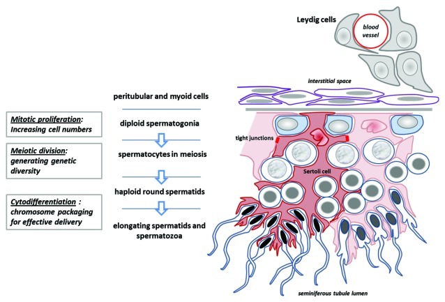
Understanding the mechanisms that lead to the differentiation of male germ cells from their spermatogonial stem cells through meiosis to give rise to mature haploid spermatozoa has been a major quest for many decades. Unlike most other cell types this differentiation process is more or less completely dependent upon the cells being located within the strongly structured niche provided by mature Sertoli cells within an intact seminiferous epithelium. While much new information is currently being obtained through the application and description of relevant gene mutations, there is still a considerable need for in vitro models with which to explore the mechanisms involved. Not only are systems of in vitro spermatogenesis important for understanding the basic science, they have marked pragmatic value in offering ex vivo systems for the artificial maturation of immature germ cells from male infertility patients, as well as providing opportunities for the transgenic manipulation of male germ cells. In this review, we have summarized literature relating to simplistic culturing of germ cells, co-cultures of germ cells with other cell types, especially with Sertoli cells, cultures of seminiferous tubule fragments, and briefly mention the opportunities of xenografting larger testicular pieces. The majority of methods are successful in allowing the differentiation of small steps in the progress of spermatogonia to spermatozoa; few tolerate the chromosomal reduction division through meiosis, and even fewer seem able to complete the complex morphogenesis which results in freely swimming spermatozoa. However, recent progress with complex culture environments, such as 3-d matrices, suggest that possibly success is now not too far away.


Figures

None
Figure 1. Cross-section of a seminiferous tubule from a mouse testis. Sertoli cells are specifically immunostained for transgenically overexpressed neurophysin. This image emphasizes clearly the different compartments (niches) in which Sertoli cells nurture specific groups of germ cells, and how the Sertoli cells effectively determine the architecture of the seminiferous epithelium.
None
Figure 2. Schematic diagram to illustrate the essential structure of the spermatogenic epithelium, its relation to the Leydig cells and interstitial space, and the manner in which the Sertoli cells determine the architecture of germ cell differentiation, as they progress from the tubule-enclosing basement membrane to the place of mature spermatozoa release in the tubule lumen (below).
None
Figure 3. Immunofluorescence microscopy to illustrate the gradual structural deterioration of the seminiferous epithelium in rat seminiferous tubules cultured in vitro for 24 h and 48 h. (A and B) are immunofluorescence images of intact cryosectioned adult rat testis, using the post-meiotic marker Dbil5 (previously endozepine-like peptide, ELP; green fluorophore). Sections were counterstained with the nuclei marker TO-PRO-3 Iodide (here red fluorophore). (Scale bar, 50µm). (C and D) represent cryosections of adult rat seminiferous tubules cultured for 24 h, as described, using the post-meiotic marker Dbil5 (red fluorophore) counterstained with DAPI (blue fluorophore), to show collapse of the tubule lumen and initial gradual mixing of different cell types within the seminiferous epithelium. (D) represents the phase-contrast image of the section in (C) in direct illumination. (E and F), as in (C and D), here cultured for 48 h. Note that now there is more substantial disruption of the epithelial structure, and the appearance of vacuoles. Nevertheless, the fact that the late spermatid marker Dbil5 is still quantitatively expressed in these tubules, shows that there has been no substantial cell death (also shown using apoptotic markers, not shown) and loss of sensitive late germ cell stages, even though there has been loss of lumen (arrows).
None
Figure 4. Intact adult rat testis (A-D) and in vitro cultured seminiferous tubules from adult rats (E-M), immunofluorescently labeled using the acrosome-specific lectin peanut agglutinin (PNA; red fluorophore) counterstained using the nuclear fluorophore DAPI (blue). (A) spermatogenic stage II-III; (B) spematogenic stage V-VI; (C) spermatogenic stage X-XI; (D) spermatogenic stage XIII-XIV. Note that the PNA highlights clearly the morphologically diverse phenotypes of the differentiating spermatids and the normal organization of the seminiferous epithelium. E-M: Cryosections from tubules cultured in vitro for 2 h (E-G), 24 h (H-J), and 48 h (K-M), as described. (E, H and K) represent so-called ‘pale’ sections of tubules, equivalent to spermatogenic stages IX to XIII; (F, I and L) correspond to ‘spot’, stages XIV to IV; (G, J and M) correspond to ‘dark’, stages VI to VIII. Note here that while there has been a degree of disruption to the organization of the seminiferous epithelium, this is not severe, although there is still loss of cells to the lumen and some disorganization of the epithelial structure.

Similar articles

Cited by

References

    1. Gilchrist RB, Thompson JG. Oocyte maturation: emerging concepts and technologies to improve developmental potential in vitro. Theriogenology. 2007;67:6–15. doi: 10.1016/j.theriogenology.2006.09.027. - DOI - PubMed
    1. Martinovitch PN. Development in vitro of the mammalian gonad. Nature. 1937;139:413. doi: 10.1038/139413a0. - DOI
    1. Sato T, Katagiri K, Gohbara A, Inoue K, Ogonuki N, Ogura A, et al. In vitro production of functional sperm in cultured neonatal mouse testes. Nature. 2011;471:504–7. doi: 10.1038/nature09850. - DOI - PubMed
    1. Ang HL, Ivell R, Walther N, Nicholson H, Ungefroren H, Millar M, et al. Over-expression of oxytocin in the testes of a transgenic mouse model. J Endocrinol. 1994;140:53–62. doi: 10.1677/joe.0.1400053. - DOI - PubMed
    1. Walker WH, Cheng J. FSH and testosterone signaling in Sertoli cells. Reproduction. 2005;130:15–28. doi: 10.1530/rep.1.00358. - DOI - PubMed

LinkOut - more resources