Review
. 2012 May;2(5):a006684.
doi: 10.1101/cshperspect.a006684.

Vascular development in the zebrafish

Affiliations
Review

Vascular development in the zebrafish

Aniket V Gore et al. Cold Spring Harb Perspect Med. 2012 May.

Abstract

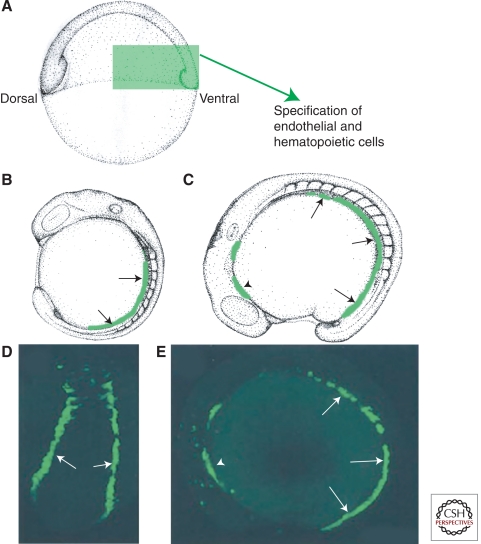
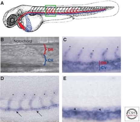
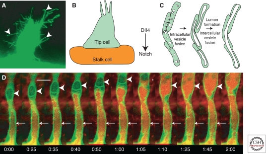
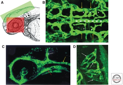
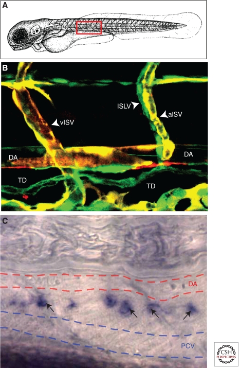
The zebrafish has emerged as an excellent vertebrate model system for studying blood and lymphatic vascular development. The small size, external and rapid development, and optical transparency of zebrafish embryos are some of the advantages the zebrafish model system offers. Multiple well-established techniques have been developed for imaging and functionally manipulating vascular tissues in zebrafish embryos, expanding on and amplifying these basic advantages and accelerating use of this model system for studying vascular development. In the past decade, studies performed using zebrafish as a model system have provided many novel insights into vascular development. In this article we discuss the amenability of this model system for studying blood vessel development and review contributions made by this system to our understanding of vascular development.


Figures

Figure 1.
Figure 1.
Endothelial cells are specified during early embryogenesis. (A) Schematic diagram of an early stage zebrafish embryo, with green boxed area indicating the region of ventral mesoderm in which endothelial precursors are specified. (B,C) Schematic diagrams of 14 somite (B) and 16 somite (C) stage embryos shown in lateral view, with migrating angioblasts shown in green. (D,E) Fluorescence micrographs of live embryos of comparable ages to B and C, with green fluorescent protein expressed under the control of an endothelial-specific promoter. Arrows in BE show migrating caudal angioblasts in the posterior lateral plate mesoderm. Arrowheads in C and E show the position of rostral angioblasts in the anterior lateral mesoderm.
Figure 2.
Figure 2.
Formation of artery and vein fates. (A) Schematic diagram of a zebrafish embryo with dorsal aorta in red and cardinal vein in blue. The green boxed area shows the approximate region in the micrographs in (BE). (B) bright-field DIC image showing position of dorsal aorta (DA) and cardinal vein (CV). Molecular markers distinguish arterial and venous endothelium even before the onset of circulation. (C) vecdn expression marks DA, CV, and intersegmental vessel (ISV) sprouts (asterisks) in 24 hpf animals. (D) dll4 is expressed in the DA (arrows) and ISV sprouts emanating from it (asterisks) but not in the CV at 24 hpf. (E) dab2 is expressed in the CV (arrowheads) but not in the DA or ISV sprouts at 24 hpf. Lateral views, anterior to the left.
Figure 3.
Figure 3.
Development of intersegmental vessels. (A) Newly formed and growing intersegmental vessel sprout with multiple cellular processes (arrowheads). (B) Schematic diagram of tip and stalk cells; Dll4 expressed in tip cells activates Notch in adjacent endothelial cells to promote stalk cell identity. (C) A model for intracellular and intercellular vesicle fusion during vascular lumenogenesis. (D) Two-photon time-lapse imaging of a lumenizing trunk intersegmental vessel in a living Tg(fli1:EGFP-cdc42wt)y48 transgenic animal injected intravascularly with 605 nm quantum dots. Quantum dots are transferred from the dorsal aorta to vacuolar compartments in proximal (arrows) and then more distal (arrowheads) intersegmental endothelial cells. In frame 1:05 the vacuole in the distal endothelial cell was captured in the process of filling with quantum dots from the proximal cell below (asterisk). For each time-lapse sequence the time from the first frame of each successive image is noted in hours:minutes. Scale bar = 20 µm. (D, From Kamei et al. 2006; reprinted, with permission, from the author.)
Figure 4.
Figure 4.
Early embryonic cranial and eye vasculature. (A) Schematic diagram of the head of a zebrafish embryo with the green and red boxed areas showing the approximate areas in the images in B, C, and D, respectively. (B) Dorsal view of the cranial vasculature showing the primordial hindbrain channels (yellow arrows), basilar artery (dashed line), and central artery sprouts (asterisks). (C) Lateral view of the cranial vasculature showing the lateral dorsal aorta (red arrow), primordial hindbrain channel (yellow arrow), and mid cerebral vessel (white arrow). (D) Dorsal view of the optic vasculature of the right eye, showing the blood vessel network surrounding the lens (white arrows). Anterior is to the left in B and C and on top in D.
Figure 5.
Figure 5.
Lymphatic vasculature in zebrafish. (A) Schematic diagram of a zebrafish embryo with a red box highlighting the approximate region shown in B and C. (B) Confocal image of blood vessels (red/yellow) and lymphatic vessels (green) in a 3 dpf Tg(flk:mCherry), Tg(fli1a:EGFP)y1 double transgenic animal. Note coalignment of intersegmental lymphatic vessels (ISLV) with arterial intersegmental vessels (aISV) but not with venous intersegmental vessels (vISV). (C) Transmitted light image of a 4 dpf zebrafish larva subjected to whole-mount in situ hybridization, probing for prox1. Expression of prox1 (black arrows) is detected in the thoracic duct (TD), a major evolutionarily conserved lymphatic vessel located between the dorsal aorta (DA, red dashed lines) and posterior cardinal vein (PCV, blue dashed lines).

References

    1. Alders M, Hogan BM, Gjini E, Salehi F, Al-Gazali L, Hennekam EA, Holmberg EE, Mannens MM, Mulder MF, Offerhaus GJ, et al. 2009. Mutations in CCBE1 cause generalized lymph vessel dysplasia in humans. Nat Genet 41: 1272–1274 - PubMed
    1. Alvarez Y, Cederlund ML, Cottell DC, Bill BR, Ekker SC, Torres-Vazquez J, Weinstein BM, Hyde DR, Vihtelic TS, Kennedy BN 2007. Genetic determinants of hyaloid and retinal vasculature in zebrafish. BMC Dev Biol 7: 114. - PMC - PubMed
    1. Alvarez Y, Astudillo O, Jensen L, Reynolds AL, Waghorne N, Brazil DP, Cao Y, O’Connor JJ, Kennedy BN 2009. Selective inhibition of retinal angiogenesis by targeting PI3 kinase. PLoS One 4: e7867. - PMC - PubMed
    1. Bayless KJ, Davis GE 2002. The Cdc42 and Rac1 GTPases are required for capillary lumen formation in three-dimensional extracellular matrices. J Cell Sci 115: 1123–1136 - PubMed
    1. Bayless KJ, Salazar R, Davis GE 2000. RGD-dependent vacuolation and lumen formation observed during endothelial cell morphogenesis in three-dimensional fibrin matrices involves the αvβ3 and α5β1 integrins. Am J Pathol 156: 1673–1683 - PMC - PubMed

Publication types

MeSH terms

Substances

LinkOut - more resources