Review
. 2012 May;2(5):a009274.
doi: 10.1101/cshperspect.a009274.

Functional neuroimaging in Parkinson's disease

Affiliations
Review

Functional neuroimaging in Parkinson's disease

Martin Niethammer et al. Cold Spring Harb Perspect Med. 2012 May.

Abstract

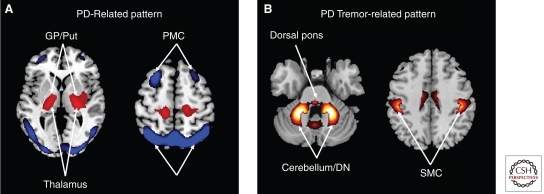
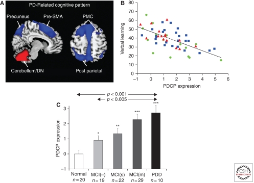
The use of functional imaging in neurodegenerative diseases has increased in recent years, with applications in research into the underlying pathophysiology, aiding in diagnosis, or evaluating new treatments. In Parkinson's disease (PD), these imaging methods have expanded our understanding of the disease beyond dopaminergic deficits. Moreover, functional imaging methods have described alterations in functional networks relating not only to the motor symptoms, but also to many nonmotor features of PD, such as cognitive dysfunction. From a clinical viewpoint, functional imaging methods can assist in monitoring disease progression, such as in the context of clinical trials, and holds the potential to aid in early diagnosis of PD and differentiation from other parkinsonian disorders.


Figures

Figure 1.
Figure 1.
Abnormal metabolic networks in Parkinson’s disease. (A) Parkinson’s disease-related pattern (PDRP). This motor-related metabolic spatial covariance pattern is characterized by hypermetabolism in the thalamus, globus pallidus (GP), pons, and primary motor cortex, associated with relative metabolic reductions in the lateral premotor (PMC) and posterior parietal areas (Ma et al. 2007). (In the representative slices, relative metabolic increases are displayed in red; relative metabolic decreases are displayed in blue. Slices were overlaid on a standard MRI brain template.) (From Eidelberg 2009; reprinted, with permission, from Elsevier © 2009.) (B) PD tremor-related metabolic pattern (PDTP) identified using a within-subject network analysis of FDG PET scans from nine tremor-dominant PD patients scanned at baseline and during ventral-intermediate (Vim) thalamic deep brain stimulation (DBS) (Mure et al. 2011). This pattern is characterized by covarying increases in the metabolic activity of the sensorimotor cortex (SMC), cerebellum, pons, and the putamen. (From Mure et al. 2011; reprinted, with permission, from Elsevier © 2011.)
Figure 2.
Figure 2.
Parkinson’s disease-related cognitive pattern: validation and correlates. (A) Parkinson’s disease-related cognitive pattern (PDCP). This cognition-related metabolic spatial covariance pattern is characterized by hypometabolism of dorsolateral prefrontal cortex (PMC), rostral supplementary motor area (preSMA), precuneus, and posterior parietal regions, associated with relative metabolic increases in the cerebellum (Huang et al. 2007a). (In the representative slices, relative metabolic increases are displayed in red; relative metabolic decreases are displayed in blue. Slices were overlaid on a standard MRI brain template.) (B) PDCP expression correlates with performance on neuropsychological tests of memory and executive functioning in nondemented PD patients. For the California Verbal Learning Test: Sum 1 to 5 (CVLT sum), this correlation was significant for the entire cohort (n = 56: r = −0.67, p < 0.001), as well as for the original group used for pattern derivation (circles, n = 15: r = −0.71, p = 0.003) and in two prospective validation groups (squares, n = 25: r = −0.53, p = 0.007; triangles, n = 16: r = −0.80, p < 0.001) (Huang et al. 2007a). (C) Bar graph of PDCP expression (mean ± SE) in PD patients with dementia (PDD), multiple-domain mild cognitive impairment (MCI[m]), single-domain mild cognitive impairment (MCI([s]), PD patients without mild cognitive impairment (MCI[−]), and in healthy control subjects. There was a significant difference in PDCP expression across the patient and control groups (F(4,70) = 8.87, p < 0.001; one-way ANOVA) and among the PD groups (F(3,56) = 4.84; p < 0.005), with higher expression in the PDD and MCI(m) cohorts compared to the MCI(−) cohort (p < 0.03; Tukey-Kramer HSD). For each PD group, PDCP expression was separately compared to healthy control values using Student's t-tests. The asterisks denote significant increases in network activity relative to controls (*p < 0.05, **p < 0.005, ***p < 0.0001) in all PD categories including MCI(−). (From Eidelberg 2009; reprinted, with permission, from Elsevier © 2009.)
Figure 3.
Figure 3.
Learning-related deactivation responses in the ventromedial prefrontal cortex (vmPFC) displayed as an inverted-U function. In addition to the influence of baseline phenotype on the cognitive response to dopaminergic therapy (Argyelan et al. 2008), we also found a significant influence of genotype. The observation that the effect of treatment on the vmPFC deactivation response varied according to COMT val158met genotype suggests that this physiologic effect is linked to intrinsic differences in prefrontal dopamine pools. Specifically, in carriers of valine alleles (VAL, i.e., valine homozygotes and val/met heterozygotes), levodopa served to suppress learning-related deactivation in this region. In contrast, recovery of this response tended to occur in individuals without this allele (MET, i.e., methionine homozygotes). The individual data were found to conform to an inverted-U function. Bands of low and high dopamine at the edges of the curve (shaded areas) represent zones of optimal function in which local deactivation responses occur during task performance. (From Argyelan et al. 2008; reprinted, with permission, from the Society for Neuroscience © 2008.)
Figure 4.
Figure 4.
Protein aggregation in Parkinson’s disease. (A) 11C-PIB uptake in a healthy volunteer, PDD subject without significant amyloid, and two DLB patients with a significant amyloid load. (From Brooks 2009; reprinted, with permission, from John Wiley & Sons © 2009.) (B) Distribution of cortical protein aggregation in regions of interest (ROIs) computed using a standard brain atlas. Binding values for [11C]-PIB and [18F]-FDDNP were estimated from PET images acquired in PD patients by measuring the target-to-cerebellum ratio >60–90 min following radiotracer injection. The values presented on the Y axis represent the difference in uptake of the two tracers in regions with metabolic reduction in the same subject.

References

    1. Aarsland D, Kurz MW 2010. The epidemiology of dementia associated with Parkinson disease. J Neurol Sci 289: 18–22 - PubMed
    1. Aarsland D, Zaccai J, Brayne C 2005. A systematic review of prevalence studies of dementia in Parkinson’s disease. Mov Disord 20: 1255–1263 - PubMed
    1. Achard S, Salvador R, Whitcher B, Suckling J, Bullmore E 2006. A resilient, low-frequency, small-world human brain functional network with highly connected association cortical hubs. J Neurosci 26: 63–72 - PMC - PubMed
    1. Adams JR, van Netten H, Schulzer M, Mak E, McKenzie J, Strongosky A, Sossi V, Ruth TJ, Lee CS, Farrer M, et al. 2005. PET in LRRK2 mutations: Comparison to sporadic Parkinson’s disease and evidence for presymptomatic compensation. Brain 128: 2777–2785 - PubMed
    1. Agdeppa ED, Kepe V, Liu J, Small GW, Huang SC, Petric A, Satyamurthy N, Barrio JR 2003. 2-Dialkylamino-6-acylmalononitrile substituted naphthalenes (DDNP analogs): Novel diagnostic and therapeutic tools in Alzheimer’s disease. Mol Imaging Biol 5: 404–417 - PubMed

Publication types

MeSH terms