

Nanoparticle-mediated p53 gene therapy for tumor inhibition
- PMID: 22553503
- PMCID: PMC3339849
- DOI: 10.1007/s13346-010-0008-9
Nanoparticle-mediated p53 gene therapy for tumor inhibition
Abstract
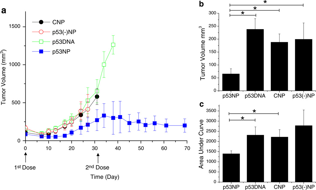
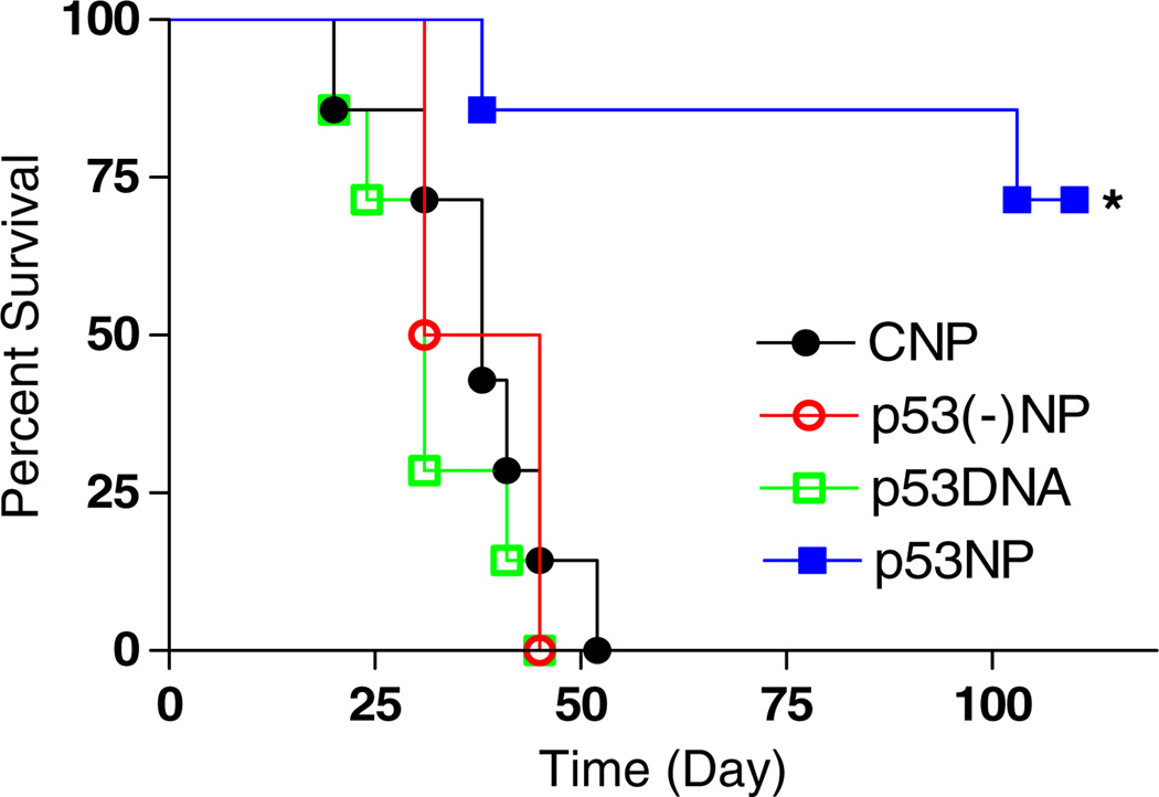
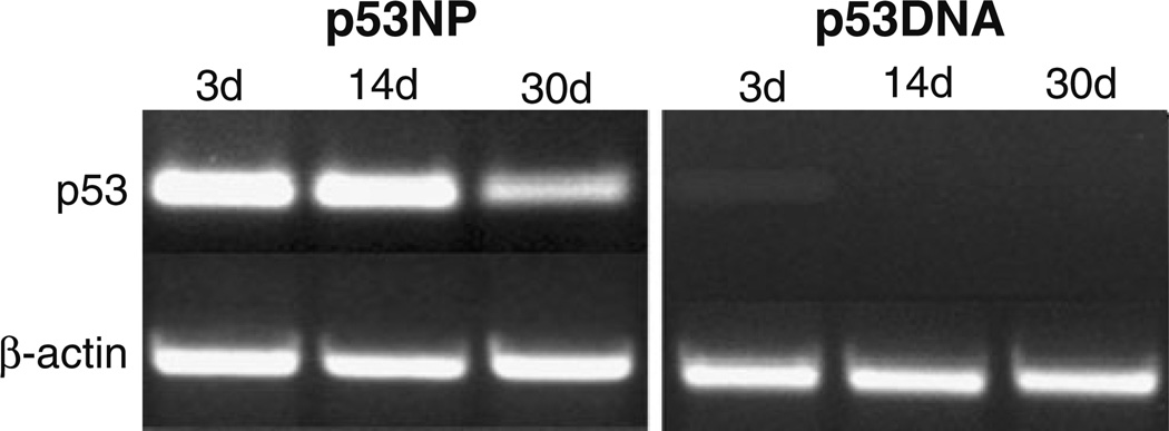
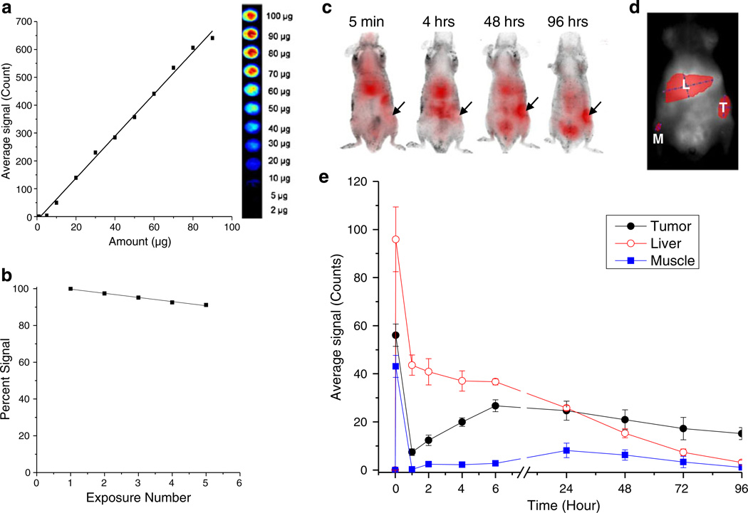
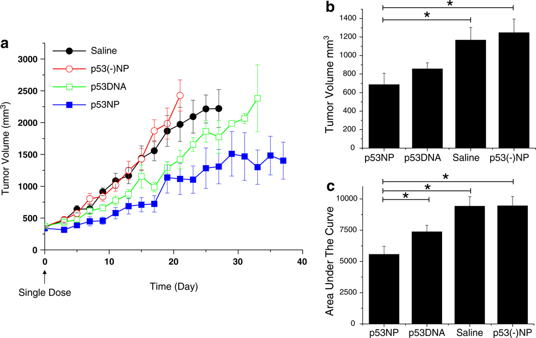
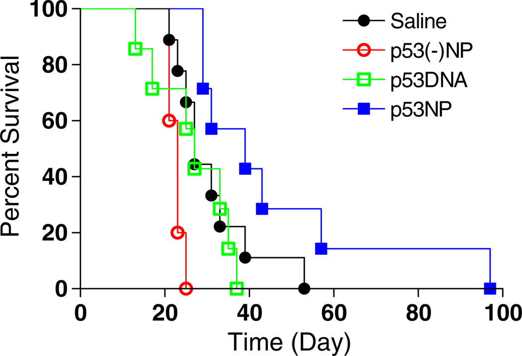
The p53 tumor suppressor gene is mutated in 50% of human cancers, resulting in more aggressive disease with greater resistance to chemotherapy and radiation therapy. Advances in gene therapy technologies offer a promising approach to restoring p53 function. We have developed polymeric nanoparticles (NPs), based on poly (lactic-co-glycolic acid), that provide sustained intracellular delivery of plasmid DNA, resulting in sustained gene expression without vector-associated toxicity. Our previous studies with p53 gene-loaded NPs (p53NPs) demonstrated sustained antiproliferative effects in cancer cells in vitro. The objective of this study was to evaluate the efficacy of p53NPs in vivo. Tumor xenografts in mice were established with human p53-null prostate cancer cells. Animals were treated with p53NPs by either local (intratumoral injection) or systemic (intravenous) administration. Controls included saline, p53 DNA alone, and control NPs. Mice treated with local injections of p53NPs demonstrated significant tumor inhibition and improved animal survival compared with controls. Tumor inhibition corresponded to sustained and greater p53 gene and protein expression in tumors treated with p53NPs than with p53 DNA alone. A single intravenous dose of p53NPs was successful in reducing tumor growth and improving animal survival, although not to the same extent as with local injections. Imaging studies showed that NPs accumulate in tumor tissue after intravenous injection; however, further improvement in tumor targeting efficiency of p53NPs may be needed for better outcome. In conclusion, the NP-mediated p53 gene therapy is effective in tumor growth inhibition. NPs may be developed as nonviral vectors for cancer and other genetic diseases.
Conflict of interest statement
Figures
References
-
- Bossi G, Sacchi A. Restoration of wild-type p53 function in human cancer: relevance for tumor therapy. Head Neck. 2007;29(3):272–284. - PubMed
-
- Bouvet M, Ellis LM, Nishizaki M, Fujiwara T, Liu W, Bucana CD, et al. Adenovirus-mediated wild-type p53 gene transfer downregulates vascular endothelial growth factor expression and inhibits angiogenesis in human colon cancer. Cancer Res. 1998;58(11):2288–2292. - PubMed
-
- Brown CJ, Lain S, Verma CS, Fersht AR, Lane DP. Awakening guardian angels: drugging the p53 pathway. Nat Rev Cancer. 2009;9(12):862–873. - PubMed
-
- Carson DA, Lois A. Cancer progression and p53. Lancet. 1995;346(8981):1009–1011. - PubMed
-
- Cicalese A, Bonizzi G, Pasi CE, Faretta M, Ronzoni S, Giulini B, et al. The tumor suppressor p53 regulates polarity of self-renewing divisions in mammary stem cells. Cell. 2009;138(6):1083–1095. - PubMed
Grants and funding
LinkOut - more resources
Full Text Sources
Research Materials
Miscellaneous
