

αB-crystallin malondialdehyde, superoxide dismutase, and lutathione peroxidase changes in X-ray irradiated rat lens
- PMID: 22553682
- PMCID: PMC3340863
- DOI: 10.3980/j.issn.2222-3959.2011.04.08
αB-crystallin malondialdehyde, superoxide dismutase, and lutathione peroxidase changes in X-ray irradiated rat lens
Abstract
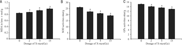
Aim: To evaluate αB-crystallin malondialdehyde (MDA), superoxide dismutase (SOD) and glutathione peroxidase (GPx) changes in X-ray irradiated rat lens.
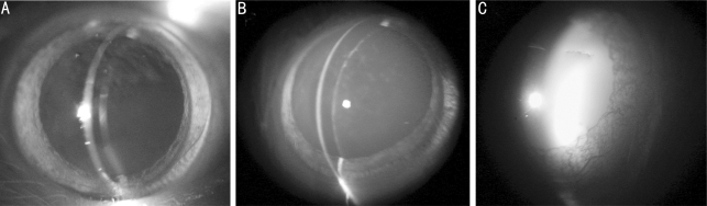
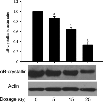
Methods: Eight-week-old Sprague-Dawley male rats received X-ray irradiation to the head with rest of the body protected. The exposure dose ranged from 2 to 25 Grays (Gy). The cataract status were examined by slit lamp and rated with "four-grade systems" post-irradiation. The lens MDA level, and the activities of SOD and GPx were measured in a short-term experiment post-irradiation, and αB-crystallin protein levels were quantified.
Results: The lenses of normal control and the X-ray irradiated groups with the dose up to 10 Gy remained transparent throughout the experiment. The lens first appeared tiny scatters, and even lamellar opacities in the posterior capsule 45 days post-irradiation with the dose of 15 Gy, and progressed slowly to the advance stage of cataract; while, for the higher dose (25 Gy), the opacity of lens appeared much earlier, and progressed more rapidly to mature stage of cataract within 1 month. At the end of the observation (90 days post-irradiation), almost all lenses became complete opacity with the higher dose (25 Gy). The degree of lens opacity was rated accordingly. The lens MDA level was increased, and SOD and GPx activities were decreased with a dose-dependent manner post-irradiation. The αB-crystallin protein level was decreased dose-dependently at the end point of observation.
Conclusion: Oxidative events and αB-crystallin may play important roles in the pathogenesis of cataract in X-ray irradiated rat lens.
Keywords: X-ray irradiation; cataract; glutathione peroxidase; superoxide dismutase; αB-crystallin malondialdehyde.
Figures
References
-
- Kyselova Z, Stefek M, Bauer V. Pharmacological prevention of diabetic cataract. J Diabetes Complications. 2004;18(2):129–140. - PubMed
-
- Toda J, Kato S, Oshika T, Sugita G. Posterior capsule opacification after combined cataract surgery and vitrectomy. J Cataract Refract Surg. 2007;33(1):104–107. - PubMed
-
- Bockelbrink A, Roll S, Ruether K, Rasch A, Greiner W, Willich SN. Cataract surgery and the development or progression of age-related macular degeneration: a systematic review. Surv Ophthalmol. 2008;53(4):359–367. - PubMed
-
- Ganea E, Harding JJ. Glutathione-related enzymes and the eye. Curr Eye Res. 2006;31(1):1–11. - PubMed
-
- Chandrasena LG, Chackrewarthy S, Perera PT, de Silva D. Erythrocyte antioxidant enzymes in patients with cataract. Ann Clin Lab Sci. 2006;36(2):201–204. - PubMed
LinkOut - more resources
Full Text Sources