

Epidemiology and molecular genetics of congenital cataracts
- PMID: 22553694
- PMCID: PMC3340856
- DOI: 10.3980/j.issn.2222-3959.2011.04.20
Epidemiology and molecular genetics of congenital cataracts
Abstract
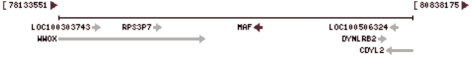
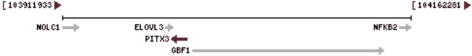
Congenital cataract is a crystallin severe blinding disease and genetic factors in disease development are important. Crystallin growth is under a combination of genes and their products in time and space to complete the coordination role of the guidance. Congenital cataract-related genes, included crystallin protein gene (CRYAA, CRYAB, CRYBA1/A3, CRYBA4, CRYBB1, CRYBB2, CRYBB3, CRYGC, CRYGD, CRYGS), gap junction channel protein gene (GJA1, GJA3, GJA8), membrane protein gene (GJA3, GJA8, MIP, LIM2), cytoskeletal protein gene (BF-SP2), transcription factor genes (HSF4, MAF, PITX3, PAX6), ferritin light chain gene (FTL), fibroblast growth factor (FGF) and so on. Currently, there are about 39 genetic loci isolated to which primary cataracts have been mapped, although the number is constantly increasing and depends to some extent on definition. We summarized the recent advances on epidemiology and genetic locations of congenital cataract in this review.
Keywords: congenital cataract; crystallin protein gene; cytoskeleton protein; ferritin light chain gene; gap junction channel protein gene; growth factor gene; membrane protein gene; transcription factor genes.
Figures


References
-
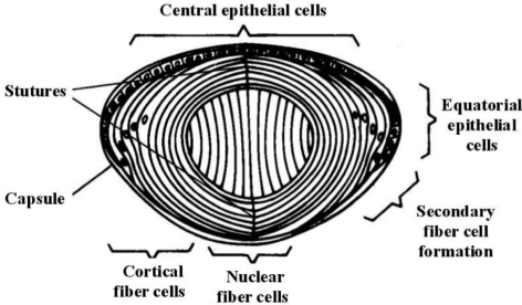
- Kuszak JR, Zoltoski RK, Sivertson C. Fibre cell organization in crystalline lenses. Exp Eye Res. 2004;78(3):673–687. - PubMed
-
- Haargaard B, Wohlfahrt J, Fledelius HC, Rosenberg T, Melbye M. A nationwide Danish study of 1027 cases of congenital/infantile cataracts: etiological and clinical classifications. Ophthalmology. 2004;111(12):2292–2298. - PubMed
-
- Johar SR, Savalia NK, Vasavada AR, Gupta PD. Epidemiology based etiological study of pediatric cataract in western India. Indian J Med Sci. 2004;58(3):115–121. - PubMed
-
- Heijl A, Leske MC. Cataract epidemiology. Ophthalmology. 2007;114(1):201. - PubMed
LinkOut - more resources
Full Text Sources
Research Materials
Miscellaneous