

Case Reports
doi: 10.3980/j.issn.2222-3959.2011.04.22.
Epub 2011 Aug 18.
Bilateral scleromalacia perforans and peripheral corneal thinning in Wegener's granulomatosis
Affiliations
- PMID: 22553696
- PMCID: PMC3340864
- DOI: 10.3980/j.issn.2222-3959.2011.04.22
Item in Clipboard
Case Reports
Bilateral scleromalacia perforans and peripheral corneal thinning in Wegener's granulomatosis
Int J Ophthalmol.
2011.
Abstract
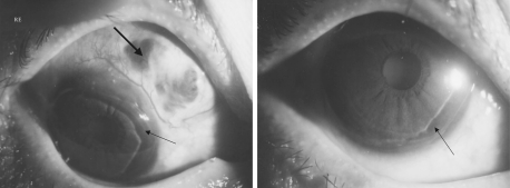
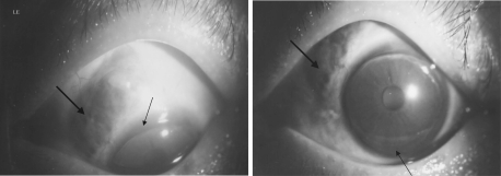
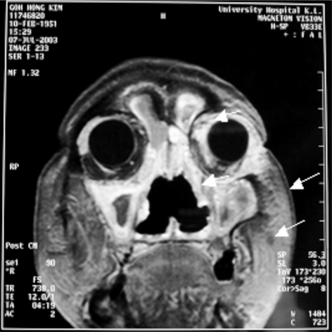
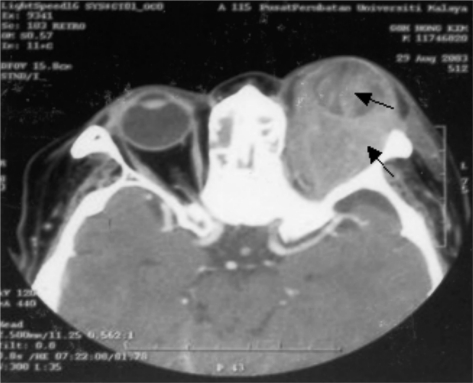
A rare case of bilateral scleromalacia perforans, bilateral peripheral corneal thinning (contact lens cornea) and unilateral orbital inflammatory disease in a 50 year old female patient with an indolent form Wegener's granulomatosis(WG) involving lungs and sinuses is reported. The patient survived for 12 years after the initial diagnosis of systemic disease. There was perforation of left globe following trauma and no perforation of the right globe till the last follow up of the patient.
Keywords: Wegener's granulomatosis; peripheral thinning of cornea; proptosis; scleromalacia perforans.
Figures
Similar articles
-
Scleromalacia perforans: a case report.J Med Case Rep. 2018 Jun 5;12(1):155. doi: 10.1186/s13256-018-1686-z. J Med Case Rep. 2018. PMID: 29866188 Free PMC article.
-
Ocular manifestations of Wegener's granulomatosis in north India.Sarcoidosis. 1988 Sep;5(2):132-5. Sarcoidosis. 1988. PMID: 3227186
-
[Atypical location of Wegener's granulomatosis with breast involvement: case report].Ann Acad Med Stetin. 2011;57(3):70-6. Ann Acad Med Stetin. 2011. PMID: 23383550 Polish.
-
Wegener's granulomatosis with orbital involvement: case report and literature review.Rom J Ophthalmol. 2021 Jan-Mar;65(1):93-97. doi: 10.22336/rjo.2021.19. Rom J Ophthalmol. 2021. PMID: 33817443 Free PMC article. Review.
-
[Wegener's and Stewart's granulomatosis: a case report of Stewart's granulomatosis].Acta Otorhinolaryngol Ital. 1998 Oct;18(5):322-31. Acta Otorhinolaryngol Ital. 1998. PMID: 10361746 Review. Italian.
Cited by
-
Systemic diseases and the cornea.Exp Eye Res. 2021 Mar;204:108455. doi: 10.1016/j.exer.2021.108455. Epub 2021 Jan 21. Exp Eye Res. 2021. PMID: 33485845 Free PMC article. Review.
-
Ocular Manifestations of Granulomatosis with Polyangiitis: A Review of the Literature.Ophthalmol Ther. 2019 Jun;8(2):227-234. doi: 10.1007/s40123-019-0176-8. Epub 2019 Mar 15. Ophthalmol Ther. 2019. PMID: 30875067 Free PMC article. Review.
-
Atypical Granulomatosis with Polyangiitis Presenting with Meibomitis, Scleritis, Uveitis and Papillary Bladder Tumor: A Case Report and Literature Review.Diagnostics (Basel). 2021 Apr 9;11(4):680. doi: 10.3390/diagnostics11040680. Diagnostics (Basel). 2021. PMID: 33918928 Free PMC article.
-
Scleromalacia perforans: a case report.J Med Case Rep. 2018 Jun 5;12(1):155. doi: 10.1186/s13256-018-1686-z. J Med Case Rep. 2018. PMID: 29866188 Free PMC article.
References
-
- Pakrou N, Selva D, Leibovitch I. Wegener's granulomatosis: Ophthlmic manifeatations and management. Semin Arthritis Rheum. 2006;35(5):284–292. - PubMed
-
- Foster CS, Yang J. Wegner's granulomatosis. In: Albert DM, Jakobiec FA, editors. Principles and practice of ophthalmology. 2nd ed. Vol. 5. Vol. 2000. Philadelphia: WB saunders; pp. 4578–4588. In:
-
- Harman LE, Margo CE. Wegner's granulomatosis. Surv Ophthalmol. 1998;42(5):458–480. - PubMed
-
- Sadiq SA, Jennings CR, Jones Ns, Downes RN. Wegener's granulomatosis: the ocular manifestations revisited. Orbit. 2000;19(4):253–261. - PubMed
-
- Bullen CL, Leisegang TJ, McDonald TJ, DeRemee RA. Ocular complications of Wegener' granulomatosis. Ophthalmology. 1983;90(3):279–290. - PubMed
Publication types
LinkOut - more resources
Full Text Sources