

Changes in endothelial cell density following penetrating keratoplasty and deep anterior lamellar keratoplasty
- PMID: 22553737
- PMCID: PMC3340783
- DOI: 10.3980/j.issn.2222-3959.2011.06.14
Changes in endothelial cell density following penetrating keratoplasty and deep anterior lamellar keratoplasty
Abstract
Aim: To compare the corneal endothelial cell density (ECD) of clear grafts after penetrating keratoplasty (PK) and deep anterior lamellar keratoplasty (DALK).
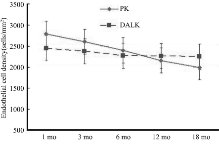
Methods: The study included 44 and 54 patients treated with PK and DALK, respectively, between March 2006 and April 2010. Corneal ECD was examined using specular microscopy at postoperative 1, 3, 6, 12, and 18 months, and the values were compared.
Results: Corneal ECD reduction in the PK group was 7.4%, 15.2%, 23.5%, and 28.9% at 3, 6, 12 and 18 months respectively after surgery, compared with 4.2 % in the first month (P<0.01). These figures were 3.0%, 6.7%, 7.2%, and 7.7% at 3, 6, 12 and 18 months respectively, compared with 2.2 % in the first month in the DALK group (P>0.05).
Conclusion: Compared with DALK, PK significantly reduced ECD of the clear grafts. These results suggest that survival of endothelial cells in grafts is better after DALK than after PK.
Keywords: endothelial cell density; lamellar keratoplasty; penetrating keratoplasty; specular microscopy.
Figures
References
-
- Bourne WM. One-year observation of transplanted human corneal endothelium. Ophthalmology. 1980;87(7):673–679. - PubMed
-
- Bourne WM. Cellular changes in transplanted human corneas, Castroviejo lecture. Cornea. 2001;20(6):560–569. - PubMed
-
- Bourne WM, Hodge DO, Nelson LR. Corneal endothelium five years after transplantation. Am J Ophthalmol. 1994;118(2):185–196. - PubMed
-
- Patel SV, Hodge DO, Bourne WM. Corneal endothelium and postoperative outcomes 15 years after penetrating keratoplasty. Am J Ophthalmol. 2005;139(2):311–319. - PubMed
-
- Melles GRJ, Remeijer L, Geerards AJM, Beekhuis WH. A quick surgical technique for deep lamellar keratoplasty using visco-dissection. Cornea. 2000;19(4):427–432. - PubMed
LinkOut - more resources
Full Text Sources
Miscellaneous