. 2012;5(1):7-12.
doi: 10.3980/j.issn.2222-3959.2012.01.02. Epub 2012 Feb 18.

Inhibited experimental corneal neovascularization by neutralizing anti-SDF-1α antibody

Affiliations

Inhibited experimental corneal neovascularization by neutralizing anti-SDF-1α antibody

Gao-Qin Liu et al. Int J Ophthalmol. 2012.

Abstract

Aim: To explore the effect of SDF-1α on the development of experimental corneal neovascularization (CRNV).

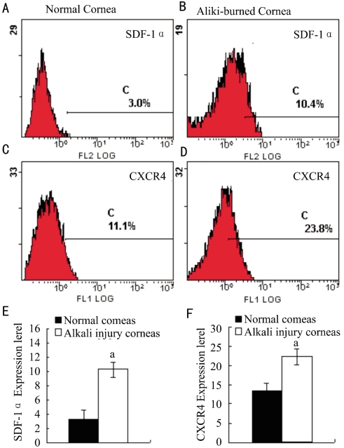
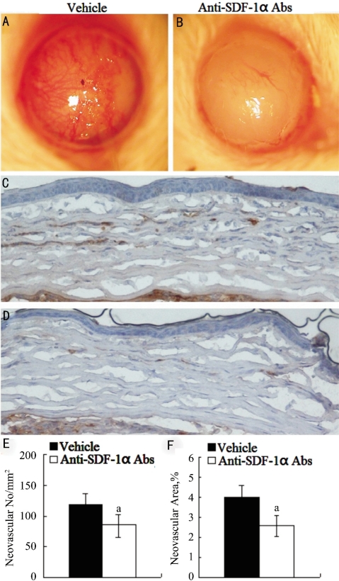
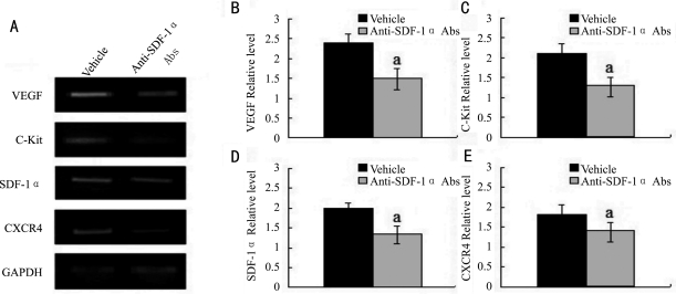
Methods: CRNV was induced by alkali injury in mice. The expression of SDF-1α and CXCR4 in burned corneas was examined by Flow Cytometry. Neutralizing anti-mouse SDF-1α antibody was locally administrated after alkali injury and the formation of CRNV 2 weeks after injury was assessed by Immunohistochemistry. The expression of VEGF and C-Kit in burned corneas was detected by RT-PCR.

Results: The number of CRNV peaks at 2 weeks after alkali injury. Compared to control group, SDF-1α neutralizing antibody treatment significantly decreased the number of CRNV. RT-PCR confirmed that SDF-1α neutralizing antibody treatment resulted in decreased intracorneal VEGF and C-Kit expression.

Conclusion: SDF-1α neutralizing antibody treated mice exhibited impaired experimental CRNV through down regulated VEGF and C-Kit expression.

Keywords: alkali injury; chemokine; corneal neovascularization.


Figures

Figure 1
Figure 1. SDF-1α and CXCR4 protein expression in normal and alkali injured corneas.
Figure 2
Figure 2. Alkali injury-induced CRNV in anti-SDF-1α mAbs treated and vehicle treated BALB/c mice
A, B: Macroscopic appearance of eyes from vehicle and anti-SDF-1α mAbs treated BALB/c mice 2 weeks after alkali injury; C, D: Tissues were immunostained with anti-CD31 antibody, and representative results are shown here. Original magnifications×400; E: CRNV numbers per square millimeter in whole section; F: Percentage CRNV areas in hot spots were determined from the corneas obtained from vehicle and anti-SDF-1α mAbs treated BABL/c mice 2 weeks after injury. Each value represents mean±SEM (n=5-8 animals). aP<0.05 vs vehicle-treated mice.
Figure 3
Figure 3. RT-PCR analysis of gene expression in the injured corneas of vehicle and anti-SDF-1α mAbs treated BALB/c mice
A: Representative result from three independent experiments of RT-PCR; B-E: Ratios of indicated genes to GAPDH of alkali-injured vehicle (black bar) and anti-SDF-1α mAbs treated BALB/c mice (grey bar) determined by RT-PCR at the indicated time intervals after alkali injury. All values represent mean±SEM (n=5-8 animals). aP<0.05 vs vehicle-treated mice.

Similar articles

Cited by

References

    1. Ambati BK, Nozaki M, Singh N, Takeda A, Jani PD, Suthar T, Albuquerque RJ, Richter E, Sakurai E, Newcomb MT, Kleinman ME, Caldwell RB, Lin Q, Ogura Y, Orecchia A, Samuelson DA, Agnew DW, St Leger J, Green WR, Mahasreshti PJ, Curiel DT, Kwan D, Marsh H, Ikeda S, Leiper LJ, Collinson JM, Bogdanovich S, Khurana TS, Shibuya M, Baldwin ME, Ferrara N, Gerber HP, De Falco S, Witta J, Baffi JZ, Raisler BJ, Ambati J. Corneal avascularity is due to soluble VEGF receptor-1. Nature. 2006;443:993–997. - PMC - PubMed
    1. Zhang S, Ma J. Ocular neovascularization: Implication of endogenous angiogenic inhibitors and potential therapy. Prog Retin Eye Res. 2007;26:1–37. - PubMed
    1. Aiuti A, Webb IJ, Bleul C, Springer T, Gutierrez-Ramos JC. The chemokine SDF-1 is a chemoattractant for human CD34+ progenitor cells and provides a new mechanism to explain the mobilization of CD34+ progenitors to peripheral blood. J Exp Med. 1997;185:111–120. - PMC - PubMed
    1. Bleul CC, Fuhlbrigge RC, Casasnovas JM, Aiuti A, Springer TA. A highly efficacious lymphocyte chemoattractant, stromal cell-derived factor 1 (SDF-1) J Exp Med. 1996;184:1101–1109. - PMC - PubMed
    1. Pablos JL, Amara A, Bouloc A, Santiago B, Caruz A, Galindo M, Delaunay T, Virelizier JL, Arenzana-Seisdedos F. Stromal-cell derived factor is expressed by dendritic cells and endothelium in human skin. Am J Pathol. 1999;155:1577–1586. - PMC - PubMed

LinkOut - more resources