

Auto-cortex of crystalline lens-induced iris neovascularization
- PMID: 22553749
- PMCID: PMC3340841
- DOI: 10.3980/j.issn.2222-3959.2012.01.05
Auto-cortex of crystalline lens-induced iris neovascularization
Abstract
Aim: To investigate auto-cortex of crystalline lens induced iris neovascularization (INV).
Methods: Thirty-six eyes of 36 guinea-pigs were included and divided into three groups randomly in this cohort study. Group A: the right lens nucleus was extracted and the remaining cortical lens material was aspirated thoroughly. Group B: the lens was removed and 30µL precipitated lens cortex was injected into the anterior chamber again. Group C: aspirated the lens cortex of the left eyes and inject them into the right anterior chambers about 10µL. Clinical changes were followed by slit-lamp examination and photograph. The eye balls were enucleated at the day of 2, 4, 7, 11, 13, 17 after operation. HE was used to detect the pathological changes.
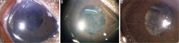
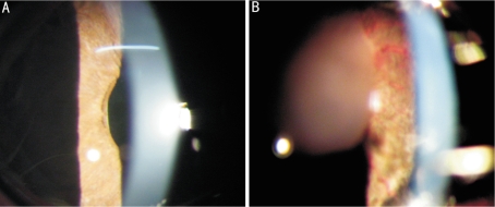
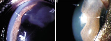
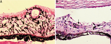
Group a: INV had not been observed until the end of empirical study. The stromal layer contained thick wall vessels, without expansion. Group B: All eyes developed INV. Postoperative (po) 7 days; the eyes developed intense and extensive INV. The vessels of iris expanded remarkably and neovascularization was observed erupting from it's lateral wall and stretching towards the anterior surface. Po11 days, INV regressed gradually after lens cortex had been absorbed. Group C: Po four (4) days, new blood vessels liking red line were presented on the anterior surface of the iris and they were not obvious.
Conclusion: Anterior chamber inside lens coriaceous can induce iris new blood vessels.
Keywords: crystallins; iris; new vessels.
Figures
References
-
- Umeda N, Ozaki H, Hayashi H, Kondo H, Uchida H, Oshima K. Non-paralleled increase of hepatocyte growth factor and vascular endothelial growth factor in the eyes with angiogenic and nonangiogenic fibroproliferation. Ophthalmic Res. 2002;34:43–47. - PubMed
-
- Zhou XL, Li YP, Zhang WX, Li ZR. Epithelial-smooth muscle-like cell differentiation of lens epithelium after micro-trauma of mouse lens. Chin Ophthalmic Res. 2007;25:721–724.
-
- Zhou HY, Zhang F, Gao LQ, Yan W, Xiong Y. Photodynamic therapy of experimental INA using hematoporphyrin monomethyl ether. Chin Ophthalmic Res. 2005;23:617–620.
-
- Shabo AL, Maxwell DS, Shintaku P, Kreiger AE, Straatasma BR. Experimental immunogenic rubeosis iridis. Invest Ophthalmol Vis Sci. 1977;16:343–352. - PubMed
-
- Stefansson E, Landers MB, III, Wolbarsht ML, Klintworth GK. Neovascularization of the iris: an experimental model in cats. Invest Ophthalmol Vis Sci. 1984;25:361–364. - PubMed
LinkOut - more resources
Full Text Sources