. 2012 May 3:11:49.
doi: 10.1186/1475-2840-11-49.

Ginkgo biloba extract reduces high-glucose-induced endothelial adhesion by inhibiting the redox-dependent interleukin-6 pathways

Affiliations

Ginkgo biloba extract reduces high-glucose-induced endothelial adhesion by inhibiting the redox-dependent interleukin-6 pathways

Jia-Shiong Chen et al. Cardiovasc Diabetol. .

Abstract

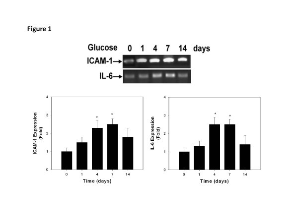
Background: Chronic elevation of glucose level activates vascular inflammation and increases endothelial adhesiveness to monocytes, an early sign of atherogenesis. This study aimed to elucidate the detailed mechanisms of high-glucose-induced endothelial inflammation, and to investigate the potential effects of Ginkgo biloba extract (GBE), an antioxidant herbal medicine, on such inflammation.

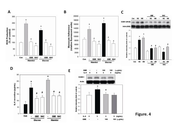
Materials and methods: Human aortic endothelial cells were cultured in high glucose or mannitol as osmotic control for 4 days. The expression of cytokines and adhesion molecules and the adhesiveness of endothelial cells to monocytes were examined. The effects of pretreatment of GBE or N-acetylcysteine, an antioxidant, were also investigated.

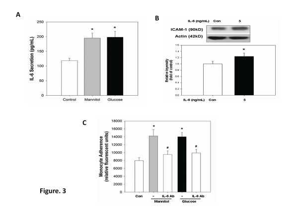
Results: Either high glucose or mannitol significantly increased reactive oxygen species (ROS) production, interleukin-6 secretion, intercellular adhesion molecule-1 (ICAM-1) expression, as well as endothelial adhesiveness to monocytes. The high-glucose-induced endothelial adhesiveness was significantly reduced either by an anti-ICAM-1 antibody or by an interleukin-6 neutralizing antibody. Interleukin-6 (5 ng/ml) significantly increased endothelial ICAM-1 expression. Piceatannol, a signal transducer and activator of transcription (STAT) 1/3 inhibitor, but not fludarabine, a STAT1 inhibitor, suppressed high-glucose-induced ICAM-1 expression. Pretreatment with GBE or N-acetylcysteine inhibited high-glucose-induced ROS, interleukin-6 production, STAT1/3 activation, ICAM-1 expression, and endothelial adhesiveness to monocytes.

Conclusions: Long-term presence of high glucose induced STAT3 mediated ICAM-1 dependent endothelial adhesiveness to monocytes via the osmotic-related redox-dependent interleukin-6 pathways. GBE reduced high-glucose-induced endothelial inflammation mainly by inhibiting interleukin-6 activation. Future study is indicated to validate the antioxidant/anti-inflammatory strategy targeting on interleukin-6 for endothelial protection in in vivo and clinical hyperglycemia.


Figures

Figure 1
Figure 1
Time-dependent effects of high glucose (D-glucose, 25 mmol/l) on ICAM-1 & IL-6 expression in human aortic endothelial cells. Western blotting analyses (upper panel) showed that both ICAM-1 (left lower panel) and IL-6 expression (right lower panel) were significantly increased after high glucose treatment for 4 days through 7 days and reduced after 14 days. N = 4 in each set of experiment. * p < 0.05 compared to day 0.
Figure 2
Figure 2
Cell viability reduction, ICAM-1 expression, and monocyte–endothelial cell adhesion mediated by high glucose in HAECs. (A) HAECs were treated with d-glucose (25 mM) or osmotic effect control mannitol (20 mM) for 4 days. (B) HAECs were incubated in glucose for 0, 1, 4, and 7 days. Cell lysates of ICAM-1 were determined by western blotting. (C) THP-1 was labeled with BCECF-AM and adhered to HAECs in response to normal-glucose, high-glucose, and mannitol treatment for 4 days. (D) HAECs in response to high-glucose and mannitol treatment for 4 days followed by neutralization with ICAM-1 antibody for 30 min (5 μg/ml). N = 8 in each set of experiment. *p < 0.05 compared to control group; #p < 0.05 compared to high-glucose or mannitol groups.
Figure 3
Figure 3
IL-6 production contributes to glucose-induced monocyte adhesiveness to HAECs. (A) HAECs were responsive to high glucose and mannitol for 4 days. The conditioned medium was isolated, and IL-6 expression was determined by ELISA. (B) HAECs were incubated with 5 ng/ml of IL-6 cytokine for 24 h. The presence of ICAM-1 in cell lysates was determined by western blotting. (C) HAECs were responsive to high glucose and mannitol for 3 days, followed by treatment with IL-6 antibody for 1 day (5 μg/ml). N = 6 in each set of experiment. *p < 0.05 compared to control group; #p < 0.05 compared to high-glucose or mannitol groups.
Figure 4
Figure 4
GBE inhibits high-glucose-induced ROS generation, IL-6 and ICAM-1 expression, and monocyte adhesiveness to HAECs. (A) HAECs were incubated in high glucose or mannitol for 3 days, followed by incubation with GBE (100 μg/ml) or NAC (1 mmol/l), an antioxidant agent, for 1 day. (B) THP-1 labeled with BCECF-AM adhered to HAECs in response to high-glucose or mannitol treatment for 3 days, followed by addition of GBE or NAC for 1 day. (C) HAECs were incubated in high glucose or mannitol for 3 days, followed by incubation in GBE and NAC for 1 day. (D) HAECs were incubated in glucose or mannitol for 3 days, followed by incubation with GBE or NAC for 1 day. Human IL-6 secreted into the medium was measured using ELISA. (E) HAECs were incubated with GBE for 30 min, followed by treatment with IL-6 (5 ng/ml) cytokine for 24 h. The presence of ICAM-1 was determined by western blotting. N = 6 in each set of experiment. *p < 0.05 compared to control group; #p < 0.05 compared to high-glucose or mannitol groups.
Figure 5
Figure 5
GBE inhibits high-glucose-induced activation of STAT1 and STAT3 as well as ICAM-1 accumulation in HAECs. (A) HAECs were incubated in high glucose for 3 days, followed by incubation with GBE, fludarabine (STAT1 inhibitor;20 μmol/l), or piceatannol (10 μmol/l) for 1 day. (B) HAECs were incubated in high glucose for 3 days, followed by incubation with GBE, fludarabine, piceatannol, fludarabine plus GBE, or piceatannol plus GBE for 1 day. (C and D) EMSAs for STAT1 and STAT3 were performed using nuclear extracts from human aortic endothelial cells that were cultured in high glucose or mannitol for 3 days, followed by treatment with GBE. Quantification of STAT1 and STAT3 activation in HAECs treated with high glucose combined with GBE is also shown. N = 6 in each set of experiment. *p < 0.05 compared to control group; # p < 0.05 compared to the high-glucose or mannitol groups.

Similar articles

Cited by

References

    1. Ross R. The pathogenesis of atherosclerosis: a perspective for the 1990s. Nature. 1993;362(6423):801–809. doi: 10.1038/362801a0. - DOI - PubMed
    1. Ceriello A. Postprandial hyperglycemia and diabetes complications: is it time to treat? Diabetes. 2005;54(1):1–7. doi: 10.2337/diabetes.54.1.1. - DOI - PubMed
    1. Kado S, Wakatsuki T, Yamamoto M, Nagata N. Expression of intercellular adhesion molecule-1 induced by high glucose concentrations in human aortic endothelial cells. Life Sci. 2001;68(7):727–737. doi: 10.1016/S0024-3205(00)00968-1. - DOI - PubMed
    1. Hodge DR, Hurt EM, Farrar WL. The role of IL-6 and STAT3 in inflammation and cancer. Eur J Cancer. 2005;41(16):2502–2512. doi: 10.1016/j.ejca.2005.08.016. - DOI - PubMed
    1. Morigi M, Angioletti S, Imberti B, Donadelli R, Micheletti G, Figliuzzi M, Remuzzi A, Zoja C, Remuzzi G. Leukocyte-endothelial interaction is augmented by high glucose concentrations and hyperglycemia in a NF-kB-dependent fashion. J Clin Invest. 1998;101(9):1905–1915. doi: 10.1172/JCI656. - DOI - PMC - PubMed

Publication types

MeSH terms