. 2012 Aug;65(1):60-7.
doi: 10.1016/j.cryobiol.2012.04.003. Epub 2012 Apr 23.

Ezrin functionality and hypothermic preservation injury in LLC-PK1 cells

Affiliations

Ezrin functionality and hypothermic preservation injury in LLC-PK1 cells

Tao Tian et al. Cryobiology. 2012 Aug.

Abstract

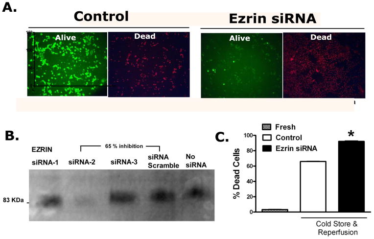
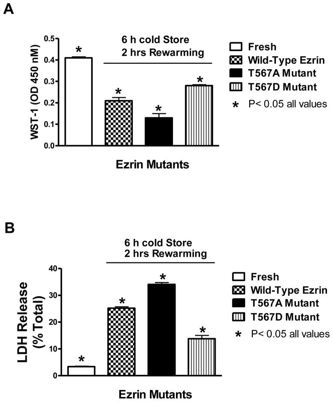
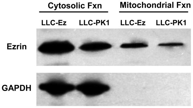
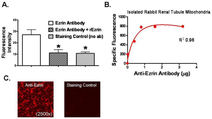
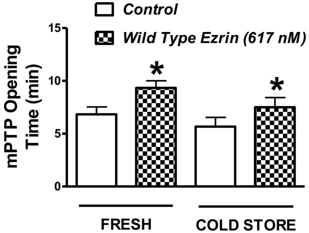
Renal epithelial cells from donor kidneys are susceptible to hypothermic preservation injury, which is attenuated when they over express the cytoskeletal linker protein ezrin. This study was designed to characterize the mechanisms of this protection. Renal epithelial cell lines were created from LLC-PK1 cells, which expressed mutant forms of ezrin with site directed alterations in membrane binding functionality. The study used cells expressing wild type ezrin, T567A, and T567D ezrin point mutants. The A and D mutants have constitutively inactive and active membrane binding conformations, respectively. Cells were cold stored (4 °C) for 6-24 h and reperfused for 1h to simulate transplant preservation injury. Preservation injury was assessed by mitochondrial activity (WST-1) and LDH release. Cells expressing the active ezrin mutant (T567D) showed significantly less preservation injury compared to wild type or the inactive mutant (T567A), while ezrin-specific siRNA knockdown and the inactive mutant potentiated preservation injury. Ezrin was extracted and identified from purified mitochondria. Furthermore, isolated mitochondria specifically bound anti-ezrin antibodies, which were reversed with the addition of exogenous recombinant ezrin. Recombinant wild type ezrin significantly reduced the sensitivity of the mitochondrial permeability transition pore (mPTP) to calcium, suggesting ezrin may modify mitochondrial function. In conclusion, the cytoskeletal linker protein ezrin plays a significant role in hypothermic preservation injury in renal epithelia. The mechanisms appear dependent on the molecule's open configuration (traditional linker functionality) and possibly a novel mitochondrial specific role, which may include modulation of mPTP function or calcium sensitivity.


Figures

Figure 1
Figure 1
Ezrin specific siRNA knock down increases preservation injury induced cell death. Panel A: Shows the Control Alive and Dead LLC-PK1 cells that have been cold stored for 24 hrs and rewarmed for 1 hour with the siRNA vehicle. The same cells 48 hrs after ezrin specific siRNA knock down show a significant increase in cell death associated with preservation injury. Panel B: Construction of an siRNA duplex specific for ezrin (Invitrogen algorithm) was tested in LLC-PK1 cells using an ezrin specific western blot. Only one duplex worked well to produce a significant depletion in ezrin expression (siRNA-2). This duplex was used in all subsequent experiments. Panel C: The bar chart quantifies the fluorescence data (from panel A) from 6 independent experiments using image analysis (Image-J v 1.43u, National Institutes of Health). The data represent the mean ± SD, * P<0.05, relative to all other groups by ANOVA.
Figure 2
Figure 2
The effects of ezrin molecular configuration on preservation injury as indexed by the WST-1 assay (A) and the LDH release assay (B). LLC-PK1 cells expressing wild type ezrin, T567A ezrin mutant (inactive), or the T567D mutant (active) were cold stored for 6 hours and then rewarmed (reperfused) for 1 additional hour to induce cold storage preservation injury. The cell culture media used for this experiment did not contain either fetal calf serum proteins or phenol red indicators, since these components interfere with the WST-1 assay. Immediately before rewarming in the incubator, the cells were given WST-1 dye to convert during the reperfusion phase. After reperfusion, the dye conversion was read on a plate reader and the LDH release was determined with a colorimetric kit. Values represent mean ± SD for 6 independent experiments that were conducted in triplicate, P< 0.05 for all values.
Figure 3
Figure 3
Western blot analysis of ezrin and GAPDH from protein extracts derived from renal tubule epithelial cells (LLC-PK1) and LLC-PK1 cells transfected with the wild type ezrin gene (LLC-Ez). LLC-Ez cells constitutively over express wild-type ezrin and produced more ezrin compared to the native LLC-PK1 cells (23). Cells were fractionated into mitochondrial and non-mitochondrial pools using a mitochondrial isolation kit for cultured cells (#89874, Thermo-Fisher Scientific, Inc., Rockford, IL, USA). The non-mitochondrial pools included cytosolic and membrane fractions. Both the purified mitochondrial and the cytosolic/membrane fractions were analyzed for ezrin and GAPDH by western blot. Ezrin was identified in both mitochondrial and cytosolic/membrane pools. GAPDH, an enzyme exclusive to the cytosol, was used to verify the purity of the mitochondrial preparation since cross contamination with cytosolic or membrane fractions could account for any or all of the observed ezrin protein identification in the mitochondrial samples. The lack of any GAPDH signal in the mitochondrial extracts imply purity of the preparation and, more importantly, indicates that the ezrin detected in the mitochondrial fraction extracts derived from the mitochondria and not from cross contamination from ezrin rich membrane or cytosolic pools.
Figure 4
Figure 4
Ezrin specific immunofluorescence detected with purified mitochondria isolated from renal proximal tubules. Panel A: Immunofluorescence intensity from digital photomicrographs (1,000x) taken of isolated, purified, and paraformaldehyde fixed mitochondria stained first with anti-ezrin primary antibody (2 μg/ml), washed, and then stained with a secondary antibody labeled with Texas Red. Fluorescence microscopy used an excitation wavelength of 596 nm and a detection wavelength of 620 nm. Three experimental conditions included mitochondria stained with specific anti-ezrin antibodies alone, with anti-ezrin antibodies plus authentic recombinant ezrin (617 nM), and a staining control where no primary antibody was used. Data from Image-J software (NIH, version 1.44p) and represent mean ± SD from 6 experiments, * P< 0.05 relative to the positive control with ezrin antibody staining alone (Ezrin Antibody). Panel B: Initial experiments with isolated fixed mitochondria stained with progressively higher concentrations of primary anti-ezrin antibodies and detected with a FITC-labeled secondary antibody using a spectrofluorometric 96-well plate reader (with excitation/emission filters set at 485/325, respectively). While the mitochondrial concentrations were constant, higher antibody concentrations produced higher specific fluorescent signals until the signals saturated at a primary antibody concentration of about 2 μg/ml). Panel C: Representative fluorescent photomicrographs of purified paraformaldehyde fixed mitochondria stained with specific anti-ezrin antibodies (left) or the staining controls (right) without the primary antibodies. Secondary Texas Red labeled antibodies were used for detection, magnification was at 2,500x.
Figure 5
Figure 5
Mitochondrial permeability transition pore (mPTP) opening in response to stepwise increases in extracellular calcium in preparations of purified mouse kidney mitochondria. The extracellular calcium concentration was increased every minute and the times represent the amount of time elapsed right before the pore opens. Mitochondria were isolated from fresh kidneys and from kidneys that were flushed with UW solution and cold stored for 24 hours. The mPTP measurements were conducted with and without the addition of exogenous recombinant ezrin (617 nM) in order to test the effects of ezrin on the sensitivity of the mPTP to calcium. The mPTP studies were conducted under warm (37° C) and oxygenated (reperfusion) conditions. Values are mean ± SD from 6 independent experiments, * P< 0.05 relative to the corresponding control value (no ezrin).

Comment in

Similar articles

Cited by

References

    1. Alderliesten M, de GM, Oldenampsen J, Qin Y, Pont C, van BL, van de WB. Extracellular signal-regulated kinase activation during renal ischemia/reperfusion mediates focal adhesion dissolution and renal injury. Am J Pathol. 2007;171:452–462. - PMC - PubMed
    1. Algrain M, Turunen O, Vaheri A, Louvard D, Arpin M. Ezrin contains cytoskeleton and membrane binding domains accounting for its proposed role as a membrane-cytoskeletal linker. J Cell Biol. 1993;120:129–139. - PMC - PubMed
    1. Apodaca G. Endocytic traffic in polarized epithelial cells: role of the actin and microtubule cytoskeleton. Traffic. 2001;2:149–159. - PubMed
    1. Aufricht C, Bidmon B, Ruffingshofer D, Regele H, Herkner K, Siegel NJ, Kashgarian M, van Why SK. Ischemic conditioning prevents Na, K-ATPase dissociation from the cytoskeletal cellular fraction after repeat renal ischemia in rats. Pediatr Res. 2002;51:722–727. - PubMed
    1. Belzer FO, Southard JH. Principles of solid-organ preservation by cold storage. Transplantation. 1988;45:673–676. - PubMed

Publication types

MeSH terms

Substances

LinkOut - more resources