Review
. 2012 Sep;1823(9):1604-16.
doi: 10.1016/j.bbamcr.2012.04.008. Epub 2012 Apr 24.

Structure, function, and assembly of heme centers in mitochondrial respiratory complexes

Affiliations
Review

Structure, function, and assembly of heme centers in mitochondrial respiratory complexes

Hyung J Kim et al. Biochim Biophys Acta. 2012 Sep.

Abstract

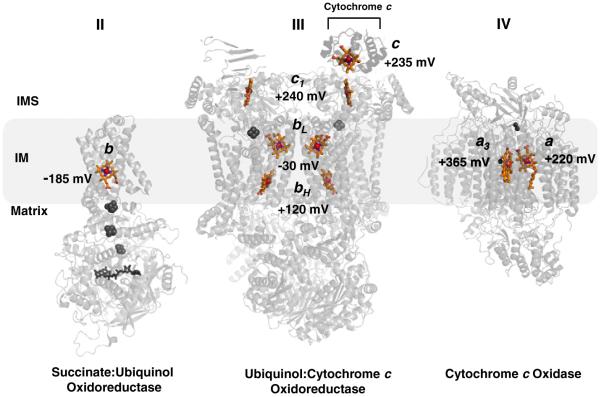
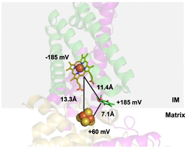
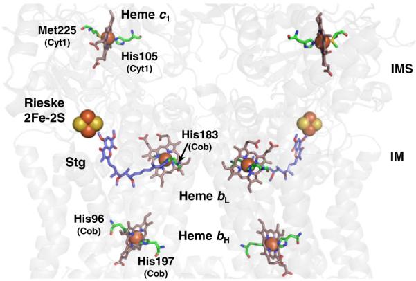
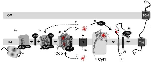
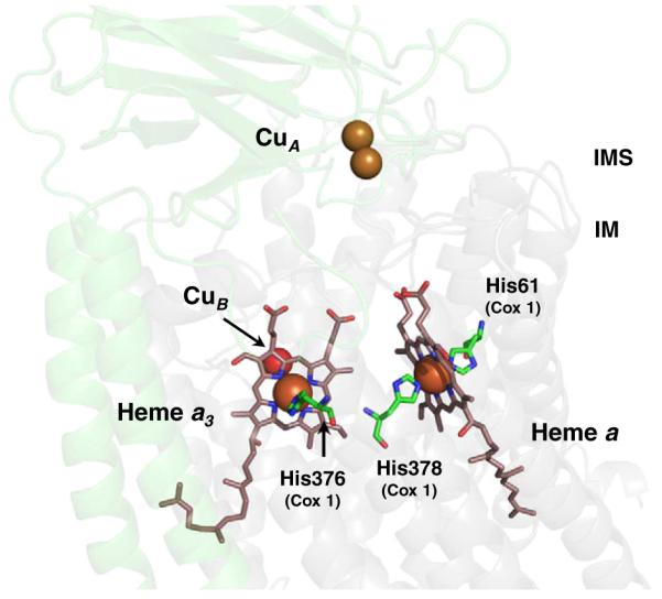
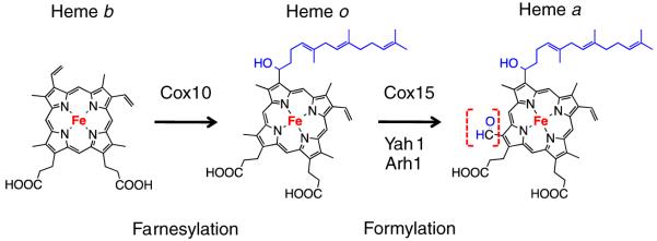
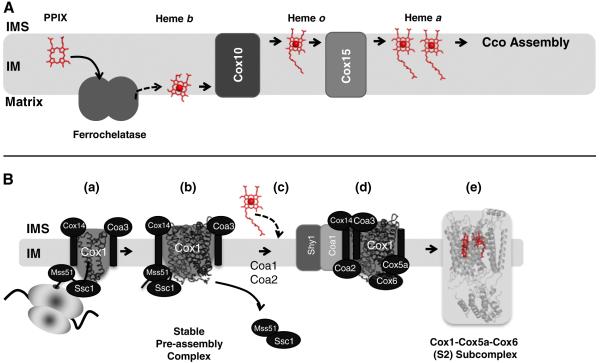
The sequential flow of electrons in the respiratory chain, from a low reduction potential substrate to O(2), is mediated by protein-bound redox cofactors. In mitochondria, hemes-together with flavin, iron-sulfur, and copper cofactors-mediate this multi-electron transfer. Hemes, in three different forms, are used as a protein-bound prosthetic group in succinate dehydrogenase (complex II), in bc(1) complex (complex III) and in cytochrome c oxidase (complex IV). The exact function of heme b in complex II is still unclear, and lags behind in operational detail that is available for the hemes of complex III and IV. The two b hemes of complex III participate in the unique bifurcation of electron flow from the oxidation of ubiquinol, while heme c of the cytochrome c subunit, Cyt1, transfers these electrons to the peripheral cytochrome c. The unique heme a(3), with Cu(B), form a catalytic site in complex IV that binds and reduces molecular oxygen. In addition to providing catalytic and electron transfer operations, hemes also serve a critical role in the assembly of these respiratory complexes, which is just beginning to be understood. In the absence of heme, the assembly of complex II is impaired, especially in mammalian cells. In complex III, a covalent attachment of the heme to apo-Cyt1 is a prerequisite for the complete assembly of bc(1), whereas in complex IV, heme a is required for the proper folding of the Cox 1 subunit and subsequent assembly. In this review, we provide further details of the aforementioned processes with respect to the hemes of the mitochondrial respiratory complexes. This article is part of a Special Issue entitled: Cell Biology of Metals.


Figures

Fig. 1
Fig. 1
Distribution of hemes b, c, and a, with respective midpoint potentials, in respiratory complexes II, III, and IV in the mitochondria. Note the general trend of increasing redox potential from the b heme of Complex II to the a3 heme of Complex IV, the site of the terminal electron acceptor and O2 reduction. Other cofactors in the complexes are grayed for clarity: Complex II; FAD, [2Fe–2S], [4Fe–4S], [3Fe–4S]: Complex III; Rieske 2Fe–2S: Complex IV; binuclear CuA, CuB. Complex III is shown as a dimer with cytochrome c docked to one of the subunit. References used for midpoint potentials: Complex II [21]; Complex III [124]; Complex IV [125]. The figure was generated using PDB accession numbers: 1YQ3, 3CX5 and 2OCC.
Fig. 2
Fig. 2
Structural and thermodynamic barriers to electron transfer from the 3Fe–4S cluster to heme b during quinone reduction in complex II. The transfer of an electron from the 3Fe–4S cluster (yellow and red spheres) to a lower potential heme b (yellow stick) would involve an “uphill” transfer, whereas a transfer to the higher potential quinone (green stick) would be more favorable thermodynamically. Furthermore, electron transfer to heme b involves a longer distance relative to the distance to the quinone. Subunit 2; yellow: Subunit 3; purple: Subunit 4; green. PDB accession number: 1YQ3 was used to generate the figure.
Fig. 3
Fig. 3
Arrangement of the hemes and the Rieske iron–sulfur clusters in the bc1 dimeric complex. For clarity, the protein backbone is in transparent grey and only the cofactors corresponding to a monomeric unit are labeled. The axial ligands to the heme iron are highlighted along with respective residue numbers and subunits (PDB ID: 3CX5 numbering). The axial ligands to heme bL are His183 and His82 (not shown) of Cob. The quinone analogue inhibitor stigmatellin (Stg) is also shown and denotes the p-side quinone-binding site that is located between the low potential b heme (Heme bL) and the Rieske iron–sulfur cluster. The n-side quinone-binding site (not shown) lies close to the high potential b heme (Heme bH).
Fig. 4
Fig. 4
Hemylation of cytochrome b and cytochrome c1 polypeptides in eukaryotic bc1 complex. Translation of COB mRNA initiates bc1 complex biogenesis (1a). Cob forms a ternary complex with the Cbp3/Cbp4/Cbp6 proteins independent of the mitochondrial ribosome (2a). Following transport of newly synthesized heme across the IM (3a), this is a possible point at which heme is inserted into the apo-cytochrome b from the IMS side of the inner membrane. This insertion could occur prior to, in conjunction with, or post Qcr7 and Qcr8 additions (4a). Transport across the IM would not be required if the product heme from ferrochelatase is deposited into the IM (see Fig. 6a). Hemylation of Cob could also occur directly from the matrix side. Apo-cytochrome c1 is synthesized in the cytosol and must be imported into the mitochondria through the inner and outer membrane translocases (1b). A bipartite signaling sequence directs insertion into the IM (2b). Following transport of newly synthesized heme across the IM, the heme lyase, Cyt2, participates in the covalent attachment of heme c1 to apocytochrome c1 (3b). The final step in holocytochrome c1 maturation is cleavage by Imp2 to remove the N-terminal membrane anchor, resulting in an Nout–Cin topology (4b). PDB accession number: 3CX5 was used to generate the figure.
Fig. 5
Fig. 5
Arrangement of the hemes a and a3:CuB and CuA in CcO. The binuclear CuA center is located in Cox2 subunit (transparent green) and is the entrance site for electrons from reduced cytochrome c. Electrons are subsequently passed to the low-spin, bis-His heme a and then to the heterobimetallic heme a3:CuB center in Cox1 (transparent grey) where O2 reduction occurs. The axial ligands to the heme iron are highlighted along with respective residue numbers and subunits (PDB ID: 2OCC numbering).
Fig. 6
Fig. 6
Chemical modifications of heme b to heme a via heme o catalyzed by the enzymes Cox10 (Heme o synthase, HOS) and Cox15 (Heme a synthase, HAS). In mitochondria, heme o is considered to be an intermediate and does not serve as a cofactor; where as in bacteria, heme o can functionally replace heme a in its terminal oxidase (e.g., cytbo3). Ferredoxin Yah1 and adrenodoxin Arh1 supply electrons for Cox15-mediated heme o to heme a conversion.
Fig. 7
Fig. 7
(A) Synthesis of heme a from protoporphyrin IX (PPIX) for hemylation of CcO in the mitochondria. PPIX (product of PPIX oxidase, not shown) likely enters ferrochelatase (shown as a dimer) from the IM. The product heme b exits either directly to the IM, or to the matrix, where it is utilized for hemylation of complex II and complex III. A sub population of heme b is converted to heme a via Cox10 and Cox15 within the IM. The exit path of heme a from Cox15 is unknown. As drawn, the heme exits on the matrix side of the IM to be used for CcO assembly. (B) Possible route of hemylation of Cox1 subunit in yeast CcO. Translation of COX1 mRNA commences CcO biogenesis (a). Newly synthesized Cox1 forms a stable complex with Mss51, Cox14, Coa3 and mitochondrial Hsp70 chaperone, Ssc1, independent of the mitochondrial ribosome (b). Subsequently, Coa1 appears to mediate progression of Cox1 to a downstream complex for cofactor insertion. Mss51 is released for further rounds of Cox1 translation/assembly (c). With the help of Shy1 and Coa2, heme a is incorporated into maturing Cox1 (d) to form the stable Cox1–Cox5a–Cox6 S2 complex (e). PDB accession number: 2OCC was used to generate the figure.

Similar articles

Cited by

References

    1. Antonini E, Brunori M. Hemoglobin and Myoglobin in their Reactions with Ligands. Elsevier; Amsterdamn and New York: 1971.
    1. Rodgers KR. Heme-based sensors in biological systems. Curr. Opin. Chem. Biol. 1999;3:158–167. - PubMed
    1. Sono M, Roach MP, Coulter ED, Dawson JH. Heme-containing oxygenases. Chem. Rev. 1996;96:2841–2888. - PubMed
    1. Mense SM, Zhang L. Heme: a versatile signaling molecule controlling the activities of diverse regulators ranging from transcription factors to MAP kinases. Cell. Res. 2006;16:681–692. - PubMed
    1. Hu R-G, Wang H, Xia Z, Varshavsky A. The N-end rule pathway is a sensor of heme. Proc. Nat. Acad. Sci. U. S. A. 2008;105:76–81. - PMC - PubMed

Publication types

MeSH terms

LinkOut - more resources