

Accuracy of linear intraoral measurements using cone beam CT and multidetector CT: a tale of two CTs
- PMID: 22554987
- PMCID: PMC3528205
- DOI: 10.1259/dmfr/21152480
Accuracy of linear intraoral measurements using cone beam CT and multidetector CT: a tale of two CTs
Erratum in
- Dentomaxillofac Radiol. 2013;42(2):20120410
Abstract
Objectives: The aim was to compare the accuracy of linear bone measurements of cone beam CT (CBCT) with multidetector CT (MDCT) and validate intraoral soft-tissue measurements in CBCT.
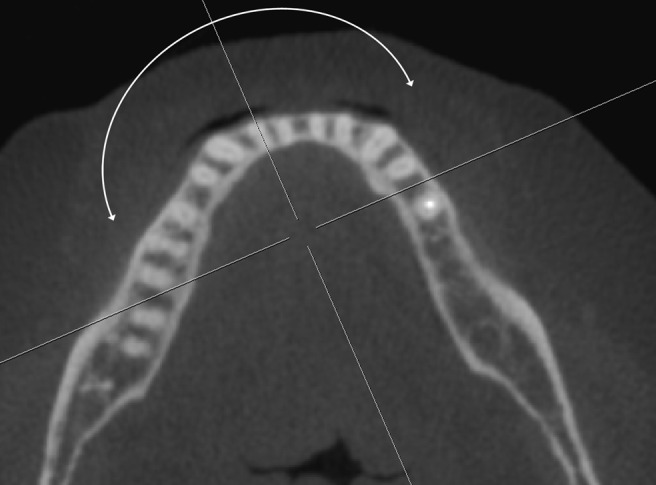
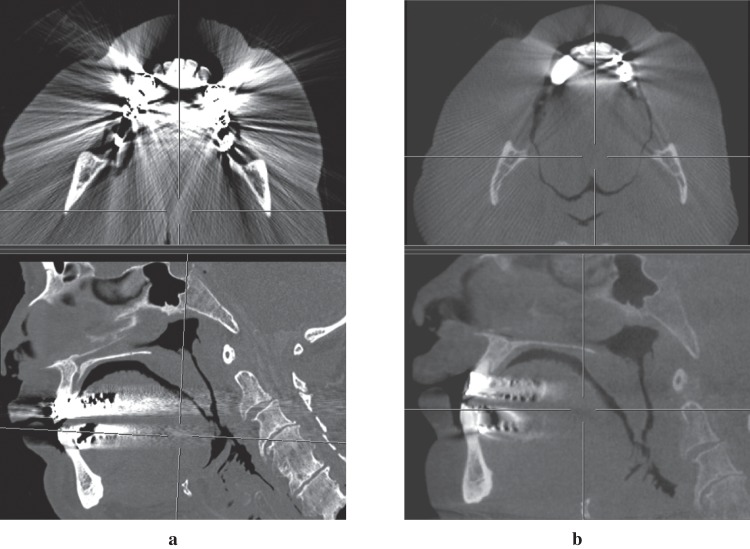
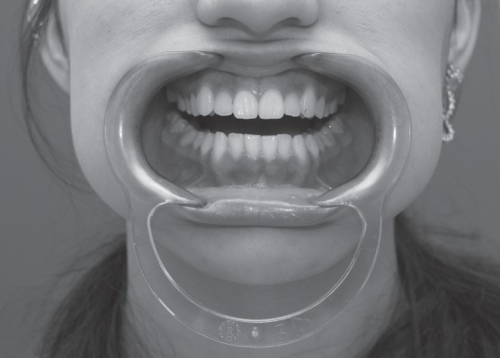
Methods: Comparable views of CBCT and MDCT were obtained from eight intact cadaveric heads. The anatomical positions of the gingival margin and the buccal alveolar bone ridge were determined. Image measurements (CBCT/MDCT) were performed upon multiplanar reformatted data sets and compared with the anatomical measurements; the number of non-assessable sites (NASs) was evaluated.
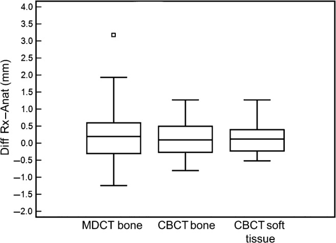
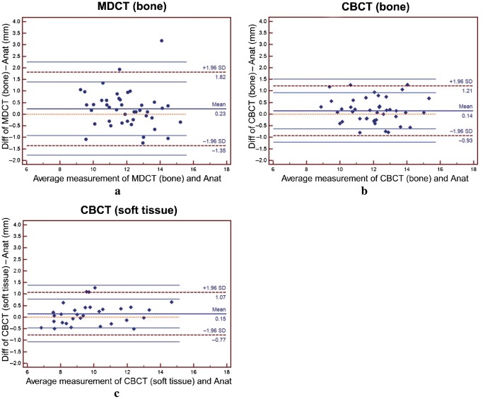
Results: Radiological measurements were accurate with a mean difference from anatomical measurements of 0.14 mm (CBCT) and 0.23 mm (MDCT). These differences were statistically not significant, but the limits of agreement for bone measurements were broader in MDCT (-1.35 mm; 1.82 mm) than in CBCT (-0.93 mm; 1.21 mm). The limits of agreement for soft-tissue measurements in CBCT were smaller (-0.77 mm; 1.07 mm), indicating a slightly higher accuracy. More NASs occurred in MDCT (14.5%) than in CBCT (8.3%).
Conclusions: CBCT is slightly more reliable for linear measurements than MDCT and less affected by metal artefacts. CBCT accuracy of linear intraoral soft-tissue measurements is similar to the accuracy of bone measurements.
Figures

Comment in
-
Accuracy of linear intraoral measurements using cone beam CT and multidetector CT: methodological mistake: author response.Dentomaxillofac Radiol. 2013;42(4):20130062. doi: 10.1259/dmfr.20130062. Epub 2013 Feb 14. Dentomaxillofac Radiol. 2013. PMID: 23412463 Free PMC article. No abstract available.
-
Accuracy of linear intraoral measurements using cone beam CT and multidetector CT: methodological mistake.Dentomaxillofac Radiol. 2013;42(4):20130048. doi: 10.1259/dmfr.20130048. Epub 2013 Feb 18. Dentomaxillofac Radiol. 2013. PMID: 23420850 Free PMC article. No abstract available.
References
-
- Robb RA, Sinak LJ, Hoffman EA, Kinsey JH, Harris LD, Ritman EL. Dynamic volume imaging of moving organs. J Med Syst 1982;6:539–554 - PubMed
-
- Arai Y, Tammisalo E, Iwai K, Hashimoto K, Shinoda K. Development of a compact computed tomographic apparatus for dental use. Dentomaxillofac Radiol 1999;28:245–248 - PubMed
-
- Hashimoto K, Arai Y, Iwai K, Araki M, Kawashima S, Terakado M. A comparison of a new limited cone beam computed tomography machine for dental use with a multidetector row helical CT machine. Oral Surg Oral Med Oral Pathol Oral Radiol Endod 2003;95:371–377 - PubMed
-
- Ludlow J, Davies-Ludlow L, Brooks S. Dosimetry of two extraoral direct digital imaging devices: NewTom cone beam CT and Orthophos Plus DS panoramic unit. Dentomaxillofac Radiol 2003;32:229–234 - PubMed
-
- Schulze D, Heiland M, Thurmann H, Adam G. Radiation exposure during midfacial imaging using 4- and 16-slice computed tomography, cone beam computed tomography systems and conventional radiography. Dentomaxillofac Radiol 2004;33:83–86 - PubMed
Publication types
MeSH terms
LinkOut - more resources
Full Text Sources
Research Materials
