

Comparison of cone beam CT device and field of view for the detection of simulated periapical bone lesions
- PMID: 22554990
- PMCID: PMC3608374
- DOI: 10.1259/dmfr/19322177
Comparison of cone beam CT device and field of view for the detection of simulated periapical bone lesions
Abstract
Objective: We aimed to assess the diagnostic accuracy of different cone beam CTs (CBCTs) and the influence of field of view (FOV) in diagnosing simulated periapical lesions.
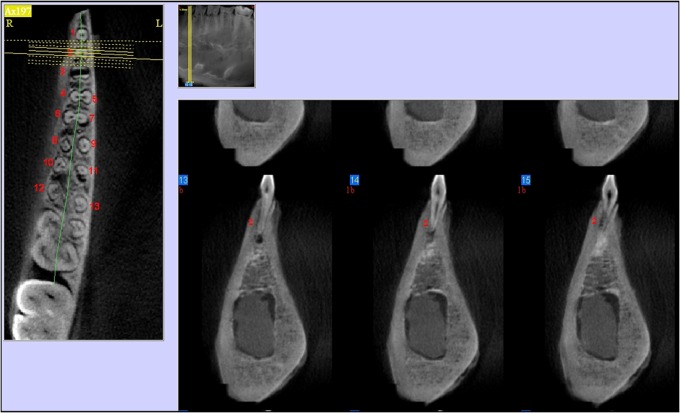
Methods: 6 formalin-fixed lateral mandibular specimens from pigs were used for creating 20 standardized periapical bone defects. 18 roots were selected for the control group. Three CBCT devices [Accuitomo 3D® (Morita, Kyoto, Japan), NewTom 3G (Quantitative Radiology, Verona, Italy) and Scanora® (Soredex, Tuusula, Finland)] and three FOVs (NewTom 3G® FOV 6, 9 and 12 inches) were used to scan the mandible. Five observers assessed the images, using a five-point probability scale for the presence of lesions. Specificity, sensitivity and areas under the receiver operating characteristic (ROC) curves were calculated.
Results: Sensitivity ranged from 72% to 80%. Specificity ranged from 60% to 77%. A difference in scoring between Scanora and the other two devices existed only in the control group. ROC analysis for different FOVs showed a decreased sensitivity with an increasing FOV, but this difference was not significant.
Conclusion: the control group, there was a difference between the CBCT devices regarding their specificity. FOV size did not show any difference in diagnostic performance. In cases in which conventional radiographic methods in combination with clinical evaluation are not sufficient, CBCT may be the method of choice to assess periapical pathology. CBCT examinations should be complementary to a clinical examination and FOV adaptation can be utilized to keep the dose to the patient as low as possible.
Figures
References
-
- Patel S, Dawood A, Whaites E, Pitt Ford T. New dimensions in endodontic imaging: part 1. Conventional and alternative radiographic systems. Int Endod J 2009;42:447–462 - PubMed
-
- Patel S. New dimensions in endodontic imaging: part 2. Cone beam computed tomography. Int Endod J 2009;42:463–475 - PubMed
-
- Van Assche N, Jacobs R, Coucke W, van Steenberghe D, Quirynen M. Radiographic detection of artificial intra-bony defects in the edentulous area. Clin Oral Implants Res 2009;20:273–279 - PubMed
-
- Lofthang-Hansen S, Huumonen S, Grondahl K. Limited cone beam computed tomography and intra-oral radiography for the diagnosis of periapical pathology. Oral Surg Oral Med Oral Pathol Oral Radiol Endod 2007;103:114–119 - PubMed
-
- Bornstein MM, Lauber R, Sendi P, von Arx T. Comparison of periapical radiography and limited cone beam computed tomography in mandibular molars for analysis of anatomical landmarks before apical surgery. J Endod 2011;37:151–157 - PubMed
Publication types
MeSH terms
LinkOut - more resources
Full Text Sources
Research Materials
