. 2012 Jul;51(3-4):347-55.
doi: 10.1016/j.molimm.2012.04.001. Epub 2012 May 1.

The specialized unfolded protein response of B lymphocytes: ATF6α-independent development of antibody-secreting B cells

Affiliations

The specialized unfolded protein response of B lymphocytes: ATF6α-independent development of antibody-secreting B cells

Ileana V Aragon et al. Mol Immunol. 2012 Jul.

Abstract

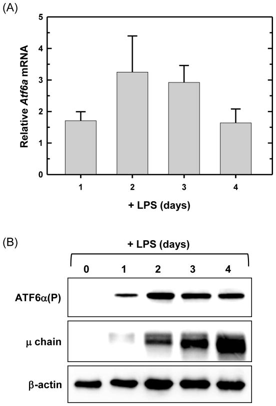
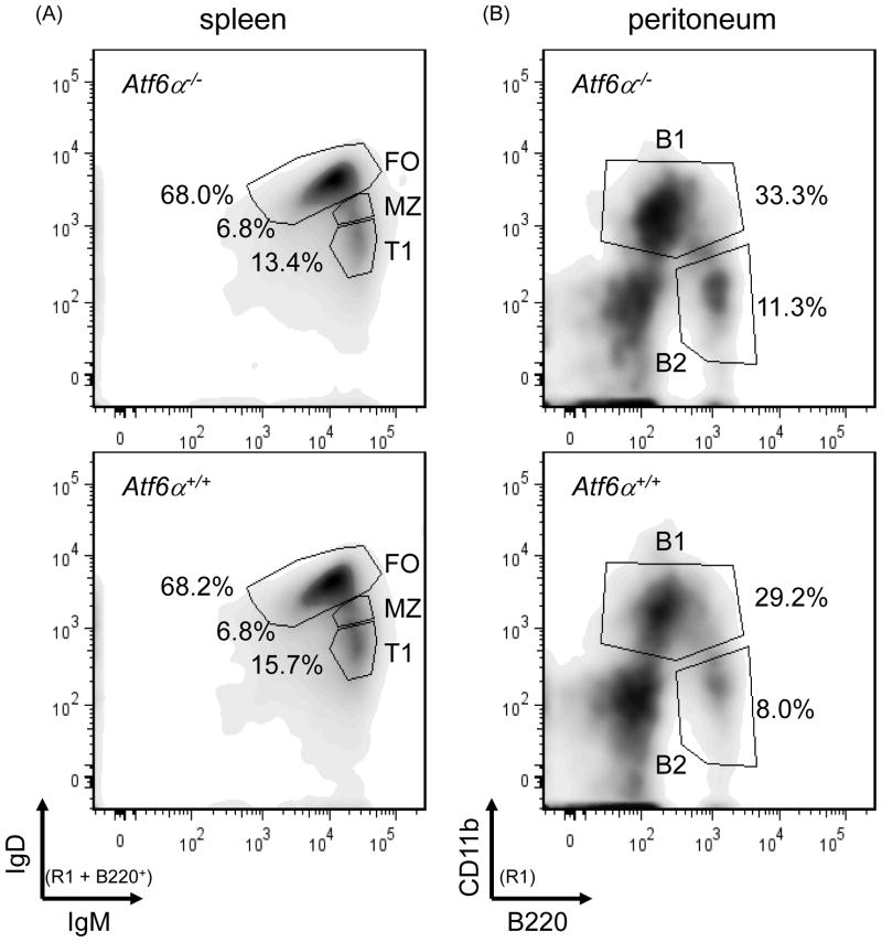
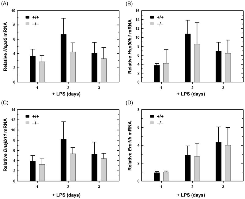
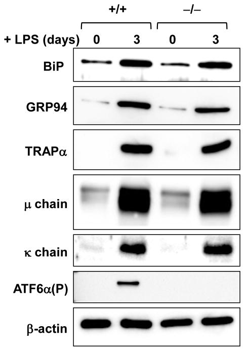
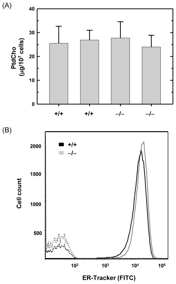
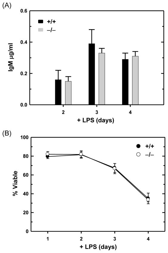
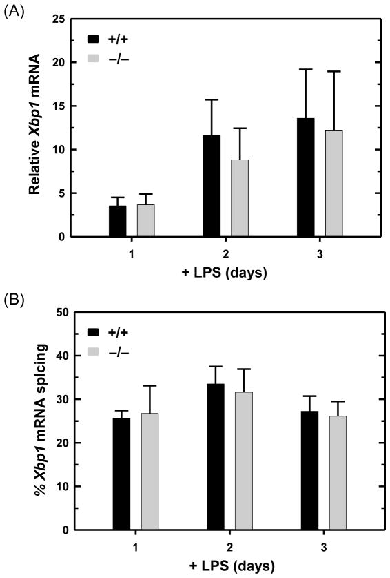
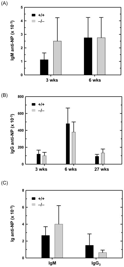
B lymphocytes, like all mammalian cells, are equipped with the unfolded protein response (UPR), a complex signaling system allowing for both pro- and mal-adaptive responses to increased demands on the endoplasmic reticulum (ER). The UPR is comprised of three signaling pathways initiated by the ER transmembrane stress sensors, IRE1α/β, PERK and ATF6α/β. Activation of IRE1 yields XBP1(S), a transcription factor that directs expansion of the ER and enhances protein biosynthetic and secretory machinery. XBP1(S) is essential for the differentiation of B lymphocytes into antibody-secreting cells. In contrast, the PERK pathway, a regulator of translation and transcription, is dispensable for the generation of antibody-secreting cells. Functioning as a transcription factor, ATF6α can augment ER quality control processes and drive ER expansion, but the potential role of this UPR pathway in activated B cells has not been investigated. Here, we report studies of ATF6α-deficient B cells demonstrating that ATF6α is not required for the development of antibody-secreting cells. Thus, when B cells are stimulated to secrete antibody, a specialized UPR relies exclusively on the IRE1-XBP1 pathway to remodel the ER and expand cellular secretory capacity.


Figures

Fig. 1
Fig. 1
Expression of ATF6α in resting and LPS-stimulated splenic B cells. Splenic B cells were isolated from wild-type mice and cultured in the presence of LPS for the indicated intervals. (A) Atf6a expression was assessed by quantitative real-time RT-PCR (qRT-PCR). Data are plotted as the level of Atf6a mRNA in LPS-stimulated cells relative to that in freshly isolated, resting cells (set at 1) (mean ± S.D., n = 3). (B) Immunoblot analysis of ATF6α(P) (full-length, precursor form), Ig μ heavy chain as a positive control for differentiation and β-actin as a control for loading and sample integrity. Data are from a representative experiment out of three repeats.
Fig. 2
Fig. 2
Flow cytometry analysis of lymphocyte populations in wild-type and ATF6α-deficient mice. Splenocytes and peritoneal cells were isolated from age- and gender-matched Atf6a+/+ and Atf6a−/− mice, stained with fluorochrome-labeled antibodies specific for cell surface markers that discriminate lymphocyte populations and analyzed by flow cytometry (R1 = lymphocyte gate as determined by visible light scatter). (A) B220+ splenocytes were assessed for levels of IgM and IgD to identify follicular (FO), marginal zone (MZ) and transitional T1 subpopulations. These subsets were further analyzed for levels of CD21, CD23, and CD93 to validate their identity and/or to identify additional subsets (Table 1 and data not shown). (B) Peritoneal cells were stained with B220 and CD11b to reveal B1 and B2 B cells. Five mice for each genotype were analyzed and representative density plots are shown.
Fig. 3
Fig. 3
Expression of ER protein folding machinery in LPS-stimulated wild-type and ATF6α-deficient B cells. Splenic B cells were isolated from Atf6a+/+ and Atf6a−/− mice and cultured in the presence of LPS for the indicated intervals. Expression of (A) Hspa5, encodes ER chaperone BiP/GRP78, (B) Hsp90b1, encodes ER chaperone GRP94, (C) Dnajb11, encodes ER chaperone co-factor ERdj3 and (D) Ero1lb, encodes ER folding enzyme ERO1-like β was assessed by qRT-PCR. Data are plotted as the level of each target mRNA in LPS-stimulated cells relative to that in freshly isolated, resting cells (set at 1) (mean ± S.D., n = 4).
Fig. 4
Fig. 4
Expression of ER proteins and Ig chains in LPS-stimulated wild-type and ATF6α-deficient B cells. Splenic B cells were isolated from Atf6a+/+ and Atf6a−/− mice and cultured in the presence of LPS for the indicated intervals. Immunoblotting was performed for the ER proteins BiP/GRP78 and GRP94, the ER translocon component TRAPα, Ig μ heavy and κ light chains, ATF6α(P) as an internal control for genotyping and β-actin as a control for loading and sample integrity. Data are from a representative experiment out of three repeats.
Fig. 5
Fig. 5
Phosphatidylcholine levels and ER abundance in LPS-stimulated wild-type and ATF6α-deficient B cells. Splenic B cells were isolated from Atf6a+/+ and Atf6a−/− mice and cultured in the presence of LPS for 3 days. (A) Lipids were extracted from equivalent numbers of cells and the amount of phosphatidylcholine (PtdCho) was determined. The experiment was performed in duplicate using B cells from two mice of each genotype; data are plotted as mean ± S.D. of triplicate determinations. (B) Cells were left unstained (hashed lines) or stained with ER-Tracker (solid lines) and analyzed by flow cytometry for ER abundance. Data are from a representative experiment out of two repeats.
Fig. 6
Fig. 6
Antibody secretion and viability of LPS-stimulated wild-type and ATF6α-deficient B cells. Splenic B cells were isolated from Atf6a+/+ and Atf6a−/− mice and cultured in the presence of LPS. (A) On the indicated days, cells were harvested, washed and replated at equivalent densities for either 2 or 3 h. Culture supernatants were then collected and assessed for IgM content by ELISA; data are plotted as the amount of IgM secreted by 5×10⁵ cells/ml/h (mean ± S.D., n = 5). (B) At the indicated intervals, cell viability was assessed by trypan blue dye exclusion (mean ± S.D., n ≥ 5 for each interval).
Fig. 7
Fig. 7
Expression and UPR-mediated splicing of Xbp1 mRNA in LPS-stimulated wild-type and ATF6α-deficient B cells. Splenic B cells were isolated from Atf6a+/+ and Atf6a−/− mice and cultured in the presence of LPS for the indicated intervals. (A) The level of total Xbp1 mRNA was assessed by qRTPCR. Data are plotted as the level of Xbp1 mRNA in LPS-stimulated cells relative to that in freshly isolated, resting cells (set at 1) (mean ± S.D., n = 3). (B) The percentage of Xbp1 transcripts modified by UPR-mediated splicing (mean ± S.D., n = 3) was determined by resolving RT-PCR products amplified from unspliced and spliced Xbp1 mRNA (237 and 211 nts, respectively) by gel electrophoresis followed by imaging and quantification.
Fig. 8
Fig. 8
Antigen-specific antibody responses in wild-type and ATF6α-deficient mice. Age- and gender-matched Atf6a+/+ and Atf6a−/− mice were immunized intraperitoneally with haptenated antigens. Blood samples were obtained at various intervals, and serial dilutions of sera were assayed by hapten-specific ELISA. Absorbance signals ≥ 2 times background (pre-immune sera) were used to determine titers of anti-NP antibodies. (A and B) For the T cell-dependent antigen NP5-KLH, mice received a primary immunization and a booster immunization 3 weeks later. Serum samples were collected at 3, 6 and 27 weeks (wks) post-primary immunization and analyzed for titers of (A) IgM and (B) IgG anti-NP antibodies (mean ± S.D., n = 4). (C) For the T cell-independent antigen NP-Ficoll, serum samples were harvested 3 weeks post-immunization and analyzed for titers of IgM and IgG3 anti-NP antibodies (mean ± S.D., n = 6).

References

    1. Adachi Y, Yamamoto K, Okada T, Yoshida H, Harada A, Mori K. ATF6 is a transcription factor specializing in the regulation of quality control proteins in the endoplasmic reticulum. Cell Struct Funct. 2008;33:75–89. - PubMed
    1. Allman D, Pillai S. Peripheral B cell subsets. Curr Opin Immunol. 2008;20:149–57. - PMC - PubMed
    1. Bommiasamy H, Back SH, Fagone P, Lee K, Meshinchi S, Vink E, Sriburi R, Frank M, Jackowski S, Kaufman RJ, Brewer JW. ATF6α induces XBP1-independent expansion of the endoplasmic reticulum. J Cell Sci. 2009;122:1626–1636. - PMC - PubMed
    1. Brunsing R, Omori SA, Weber F, Bicknell A, Friend L, Rickert R, Niwa M. B- and T-cell development both involve activity of the unfolded protein response pathway. J Biol Chem. 2008;283:17954–17961. - PubMed
    1. Calame KL, Lin KI, Tunyaplin C. Regulatory mechanisms that determine the development and function of plasma cells. Annu Rev Immunol. 2003;21:205–230. - PubMed

Publication types

MeSH terms