Review
. 2012 May;191(1):33-64.
doi: 10.1534/genetics.111.134577.

Mating-type genes and MAT switching in Saccharomyces cerevisiae

Affiliations
Review

Mating-type genes and MAT switching in Saccharomyces cerevisiae

James E Haber. Genetics. 2012 May.

Abstract

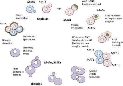
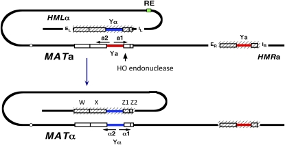
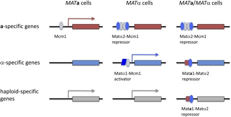
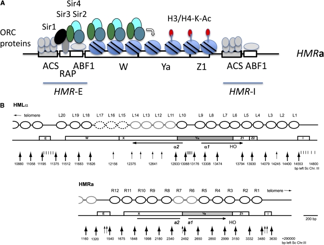
Mating type in Saccharomyces cerevisiae is determined by two nonhomologous alleles, MATa and MATα. These sequences encode regulators of the two different haploid mating types and of the diploids formed by their conjugation. Analysis of the MATa1, MATα1, and MATα2 alleles provided one of the earliest models of cell-type specification by transcriptional activators and repressors. Remarkably, homothallic yeast cells can switch their mating type as often as every generation by a highly choreographed, site-specific homologous recombination event that replaces one MAT allele with different DNA sequences encoding the opposite MAT allele. This replacement process involves the participation of two intact but unexpressed copies of mating-type information at the heterochromatic loci, HMLα and HMRa, which are located at opposite ends of the same chromosome-encoding MAT. The study of MAT switching has yielded important insights into the control of cell lineage, the silencing of gene expression, the formation of heterochromatin, and the regulation of accessibility of the donor sequences. Real-time analysis of MAT switching has provided the most detailed description of the molecular events that occur during the homologous recombinational repair of a programmed double-strand chromosome break.


Figures

Figure 1
Figure 1
Homothallic life cycle of Saccharomyce cerevisiae. A homothallic (HO) MATa (light red) mother cell and its new daughter can switch to MATα (light blue). This lineage is established by the asymmetric partitioning of the mRNA encoding the Ash1 repressor of HO gene expression in daughter cells (light green). These cells can conjugate to form a zygote that gives rise to MATa/MATα diploids (purple), in which HO gene expression is repressed. Under nitrogen starvation, diploids undergo meiosis and sporulation to produce four haploid spores (two MATa and two MATα) in an ascus. The spores germinate and grow vegetatively and can repeat the homothallic cycle. Heterothallic (ho) cells have stable mating types and grow vegetatively until they exhaust their nutrients and enter stationary phase.
Figure 2
Figure 2
Arrangement of MAT, HML, and HMR on chromosome III. The gene conversion from MATa to MATα is illustrated. Transcription of a- and α-regulatory genes at MAT are transcribed from a bidirectional promoter. Both HML and HMR could be transcribed but are silenced by the creation of short regions of heterochromatin (hatched lines) by the interaction of silencing proteins with flanking cis-acting silencer E and I sequences. The recombination enhancer (RE) located 17 kb centromere proximal to HML acts to promote the usage of HML as the donor in MATa cells.
Figure 3
Figure 3
Control of mating-type–specific genes. The Mcm1 protein, in combination with Matα1 and Matα2, activates the transcription of α-specific genes or represses a-specific genes, respectively, while a Mata1-Matα2 repressor turns off haploid-specific genes.
Figure 4
Figure 4
Silencing of HMR and HML. (A) Establishment of silencing at HMR-E. The processive process of silencing is illustrated. Proteins bound to the three elements of the HMR-E silencer recruit Sir1 that in turn recruits the Sir2-Sir3-Sir4 complex. The NAD+-dependent HDAC Sir2 deacetylates lysines on histones on the N-terminal tails of H3 and H4, which allows the Sir3-Sir4 to bind and stabilize the position of the nucleosome. Sir2 can then deactylate the next nucleosome and silencing spreads further. Here the spread of silencing is shown progressing in one direction and from one of the two silencing elements. In reality, silencing spreads from both HMR-E and HMR-I and also spreads in a limited fashion to the flanking regions. (B) Highly positioned nucleosomes in HML and HMR as determined by the Simpson lab (Weiss and Simpson 1998; Ravindra et al. 1999).
Figure 5
Figure 5
Physical monitoring of MAT switching. Southern blot analysis of StyI-digested DNA after galactose induction of HO endonuclease. The probe detects sequences just distal to MAT-Z1/Z2 and shows a difference in the size of the StyI restriction fragments of MATa and MATα. In this experiment, a ade3::GAL::HO strain carrying HMLα MATa hmrΔ cdc7-as3 was used. Cells were arrested prior to DNA replication by inhibiting Cdc7 with 1-NMPP1 (Ira et al. 2004) and then shifted to 37° to inactivate a temperature-sensitive mutation of the DNA replication factor Dpb11. In Dpb11+ cells, one can see the cleavage of MATa into a smaller HO-cut segment, followed by the appearance of the MATα product. Switching fails in absence of Dpb11 at the restrictive temperature. Data are from Hicks et al. (2011).
Figure 6
Figure 6
Mechanism of MAT switching. Key steps in the switching of MATa to MATα by a synthesis-dependent strand-annealing (SDSA) mechanism (reviewed by Pâques and Haber 1999). An HO-induced DSB is resected by 5′ to 3′ exonucleases or helicase endonucleases to produce a 3′-ended ssDNA tail, on which assembles a Rad51 filament. The Rad51::ssDNA complex engages in a search for homology. In the MAT-Z region, strand invasion can form an interwound (plectonemic) joint molecule that can assemble DNA replication factors to copy the Yα sequences. Unlike normal replication, the newly copied strand is thought to dissociate from the template and, when sufficiently extended, anneal with the second end, still blocked from forming a plectonemic structure by the long nonhomologous single-stranded Ya sequences. These sequences are clipped off once strand annealing occurs, by the Rad1-Rad10 flap endonuclease, so that the new 3′ end can be used to primer extend and copy the second strand of the Yα sequences. Consequently all newly synthesized DNA is found at the MAT locus while the donor is unaltered. A small fraction of DSB repair events apparently proceed by a different repair mechanism involving the formation of a double Holliday junction (see Pâques and Haber 1999 for details).
Figure 7
Figure 7
Detection of intermediates of MAT switching. Chromatin immunoprecipitation (ChIP) and PCR can be used to detect three early intermediates in MAT switching. (Top, from left to right) First, Rad51 assembles on the resected end of the MAT-Z region, as detected by ChIP using a pair of PCR primers specific to the MAT-distal region. Then Rad51::ssMAT-Z DNA engages the homologous sequences of HML, detected by PCR primers specific for sequences to the right of HML-Z. Finally, the initiation of new DNA synthesis is detected by a PCR assay using one primer in HML-Yα and a second primer distal to MAT-Z, so that no amplification is possible until at least 50 nt of new DNA synthesis has occurred. (Bottom) Data for these three processes are modified from Hicks et al. (2011).
Figure 8
Figure 8
Mutations arising during MAT switching. The Ya sequences of HMRa were replaced by K. lactis URA3 (Kl-URA3) sequences such that the normal HO cleavage site at the Ya-Z border was ablated (A). HML was also deleted, so that induction of HO endonuclease resulted in the switching of MATα to mat::Kl-URA3. At a rate of ∼1 in 105, the switched sequences were Ura3 and 5-FOA resistant. About half of the mutant events were single-base-pair substitutions, but the rest apparently resulted from template switching during repair, resulting in −1 frameshifts in homonucleotide runs (B), frameshifts by copying quasipalindromes (C), and interchromosomal template jumps using the homeologous ura3-52 sequences on a different chromosome (D).
Figure 9
Figure 9
Consensus elements in the RE and protein binding. (Top) DNA sequences shared by evolutionarily conserved and functional RE elements in ∼250 bp from S. cerevisiae, S. bayanus, and S. carlsbergensis. (Middle) In MATa cells, Mcm1 binding facilitates the binding of Swi4-Swi6 and multiple copies of Fkh1. (Bottom) In MATα cells, the Matα2-Mcm1 repressor binds to a 31-bp conserved operator that is shared by a-specific genes.
Figure 10
Figure 10
Role of the recombination enhancer in MATa donor preference. (A) Arrangement of HMLα, MATa, and HMRα-BamHI (HMRα-B) in wild type, REΔ, and when the RE is replaced by four LexA-binding domains to which a LexA-FHAFkh1 fusion protein can bind. (B) Southern blot data after induction of switching showing the proportion of BamHI-digested MATα or MATα-B DNA in the strains depicted above. A strain in which the LexA-FHAFkh1 domain carries a R80A mutation that prevents phosphothreonine binding fails to enhance the usage of HML (Li et al. 2012)
Figure 11
Figure 11
Model for donor preference. A cluster of Fkh1-FHA domains bound to RE in MATa cells can associate with phosphothreonine residues that are located near the DSB and created by casein kinase II, and possibly other kinases, in response to the DSB. This association tethers HMLα within ∼20 kb of the DSN ends and facilitates its use over HMR, located 100 kb away.

References

    1. Abraham J., Nasmyth K. A., Strathern J. N., Klar A. J., Hicks J. B., 1984. Regulation of mating-type information in yeast. Negative control requiring sequences both 5′ and 3′ to the regulated region. J. Mol. Biol. 176: 307–331. - PubMed
    1. Amati B. B., Gasser S. M., 1988. Chromosomal ARS and CEN elements bind specifically to the yeast nuclear scaffold. Cell 54: 967–978. - PubMed
    1. Ansari A., Gartenberg M. R., 1997. The yeast silent information regulator Sir4p anchors and partitions plasmids. Mol. Cell. Biol. 17: 7061–7068. - PMC - PubMed
    1. Arcangioli B., 1998. A site- and strand-specific DNA break confers asymmetric switching potential in fission yeast. EMBO J. 17: 4503–4510. - PMC - PubMed
    1. Arcangioli B., de Lahondes R., 2000. Fission yeast switches mating type by a replication-recombination coupled process. EMBO J. 19: 1389–1396. - PMC - PubMed

Publication types

MeSH terms