. 2012 Sep;19(9):1536-48.
doi: 10.1038/cdd.2012.46. Epub 2012 May 4.

Calpains mediate epithelial-cell death during mammary gland involution: mitochondria and lysosomal destabilization

Affiliations

Calpains mediate epithelial-cell death during mammary gland involution: mitochondria and lysosomal destabilization

T Arnandis et al. Cell Death Differ. 2012 Sep.

Abstract

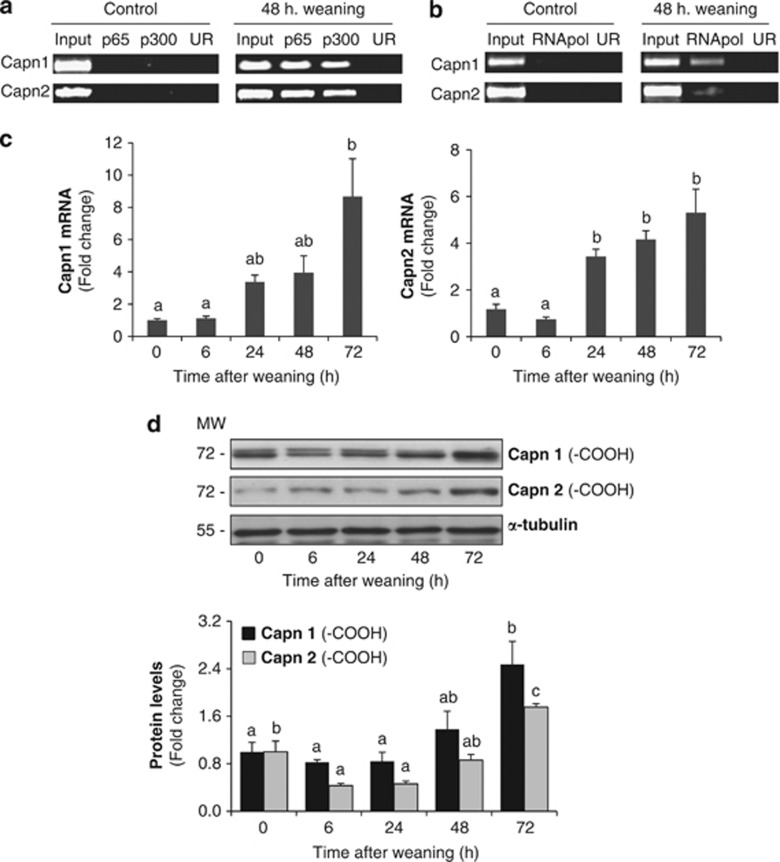
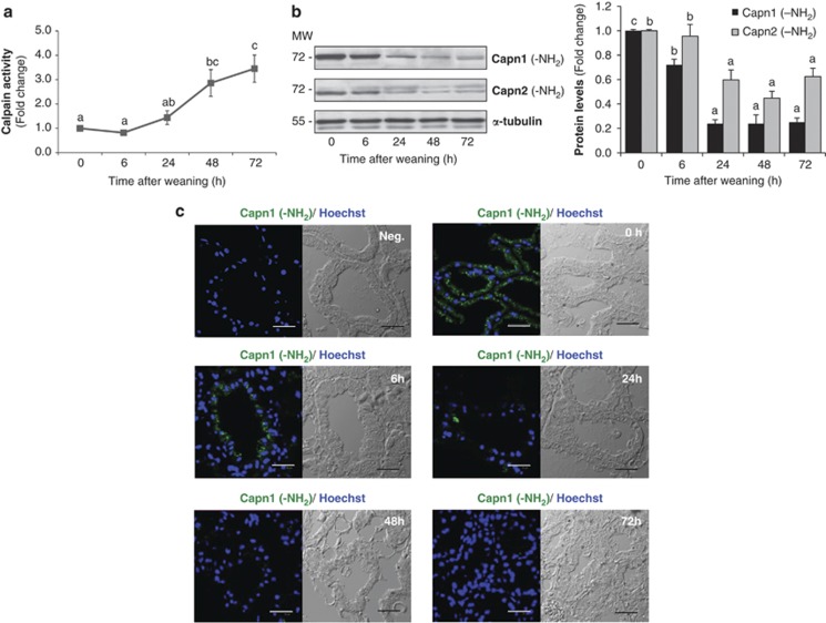
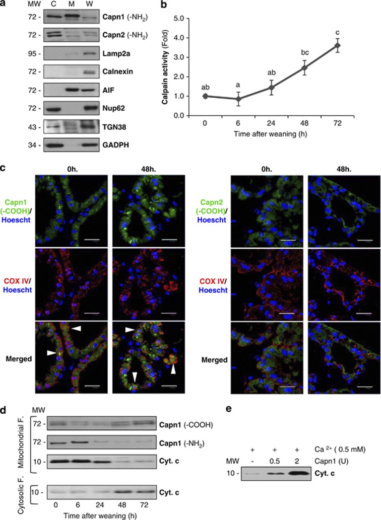
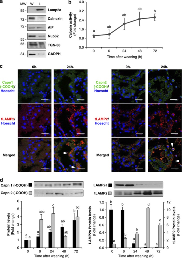
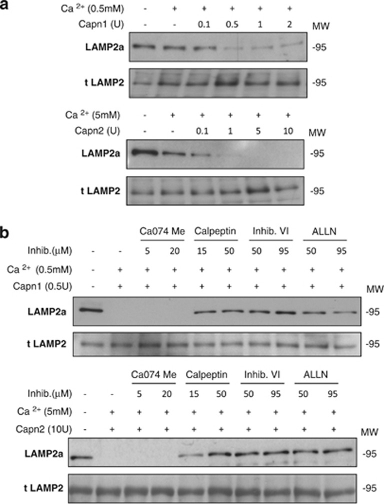
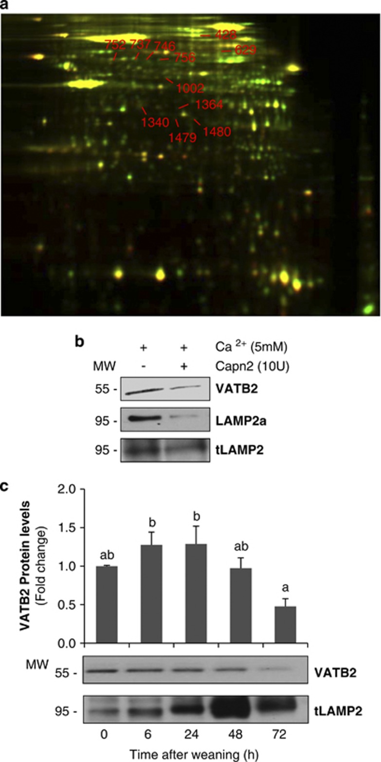
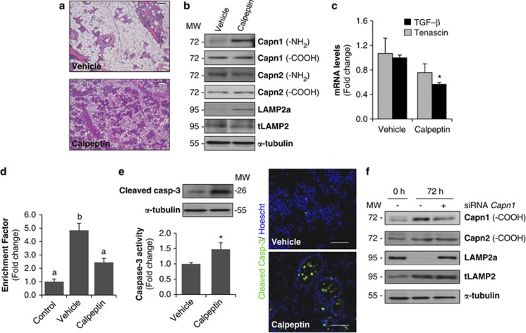
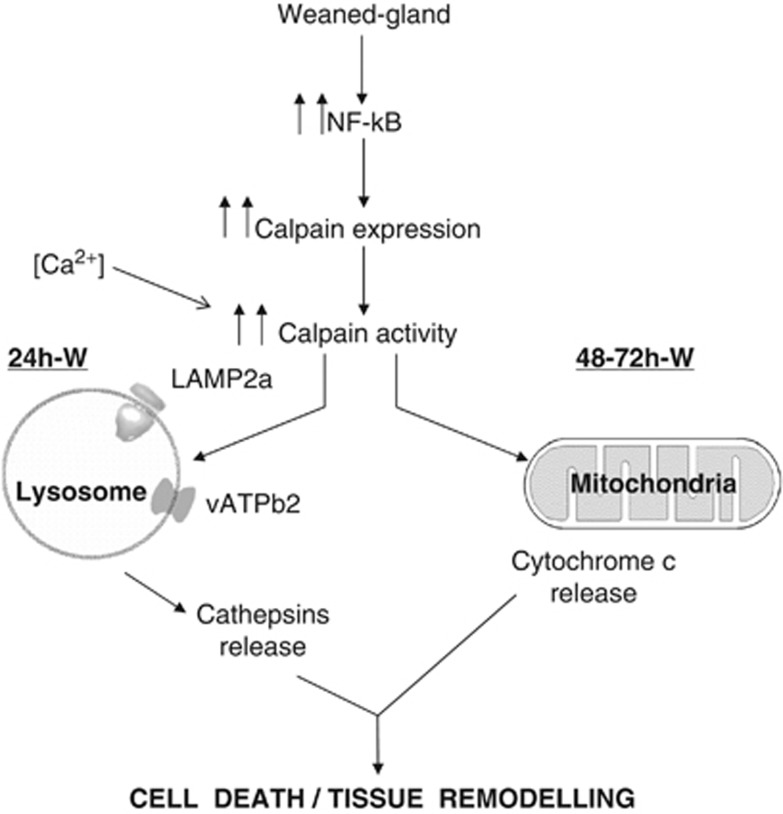
Our aim was to elucidate the physiological role of calpains (CAPN) in mammary gland involution. Both CAPN-1 and -2 were induced after weaning and its activity increased in isolated mitochondria and lysosomes. CAPN activation within the mitochondria could trigger the release of cytochrome c and other pro-apoptotic factors, whereas in lysosomes it might be essential for tissue remodeling by releasing cathepsins into the cytosol. Immunohistochemical analysis localized CAPNs mainly at the luminal side of alveoli. During weaning, CAPNs translocate to the lysosomes processing membrane proteins. To identify these substrates, lysosomal fractions were treated with recombinant CAPN and cleaved products were identified by 2D-DIGE. The subunit b(2) of the v-type H(+) ATPase is proteolyzed and so is the lysosomal-associated membrane protein 2a (LAMP2a). Both proteins are also cleaved in vivo. Furthermore, LAMP2a cleavage was confirmed in vitro by addition of CAPNs to isolated lysosomes and several CAPN inhibitors prevented it. Finally, in vivo inhibition of CAPN1 in 72-h-weaned mice decreased LAMP2a cleavage. Indeed, calpeptin-treated mice showed a substantial delay in tissue remodeling and involution of the mammary gland. These results suggest that CAPNs are responsible for mitochondrial and lysosomal membrane permeabilization, supporting the idea that lysosomal-mediated cell death is a new hallmark of mammary gland involution.


Figures

Figure 1
Figure 1
NF-κB activation regulates calpain expression during weaning. (a) Representative ChIP assay from control lactating and 48-h-weaned mammary glands showing the in vivo association of NF-κB (subunit p65) and its transcriptional coactivator p300 to the Capn1 and Capn2 promoters during weaning. (b) RNA pol II ChIP assay showing the presence of the RNApol II in the coding region of both Capn1 and Capn2. Chromatin was immunoprecipitated with anti-p65, anti-p300 or anti-RNApol II antibodies. An aliquot of the input chromatin and an unrelated antibody (UR) as negative control are also shown. Primers specific to the promoter or coding region for Capn1 and Capn2 genes were used to amplify the DNA isolated from the ChIP assay. (c) Real-time PCR was performed on both genes, Capn1 and Capn2, at different times after weaning (0, 6, 24, 48 and 72 h). Each PCR was normalized against the housekeeping gene 18S. Results are shown as mean±S.E.M. (n=3). (d) Western blot analysis for CAPN1 and CAPN2 was performed in protein mammary extracts at day 10 of lactation (0 h) and 6, 24, 48 and 72 h of involution. In CAPN1, besides the bands at 80 kDa, bands at smaller molecular weights were also detected. Three separate mice were used per developmental stage. Equal loading was confirmed by reprobing the blot against α-tubulin. The intensity of the bands was measured by densitometry using ImageJ, and quantified against α-tubulin. In (c) and (d) ANOVA was performed for the statistical analysis, where different superscript letters indicate significant differences, P<0.05; the letter ‘a' always represents the lowest value within the group
Figure 2
Figure 2
Calpain activation in mammary gland involution. (a) Protease activity was measured in mammary gland samples from control lactating (0 h) and involuting mice (6, 24, 48 and 72 h) using a fluorogenic assay based on the cleavage of the calpain substrate Suc-LLY-AFC. Results are shown as mean±S.E.M. (n=3). (b) Representative immunoblot for the amino-terminal domain of both CAPN1 and CAPN2 during involution of the mammary gland. α-Tubulin was used for normalization. In (a) and (b) ANOVA was performed for the statistical analysis, where different superscript letters indicate significant differences, P<0.05; the letter ‘a' always represents the lowest value within the group. (c) Representative immunofluorescence staining for calpain 1 amino-terminal epitope (green) in the mammary gland of control (0 h) and weaned mice (0, 6, 24, 48 and 72 h). Autofluorescence and nonspecific binding was reduced using samples incubated with dilution buffer or secondary antibody (negative control). Cell nuclei are shown in blue (Hoeschst-33342). Bright-field images were included simultaneously to visualize the exact localization of immunofluorescence in mammary gland structure. Scale bar: 30 μm
Figure 3
Figure 3
Mitochondrial effects of calpain activation. (a) Immunoblot of the cytoplasmic (C), mitochondrial (M) and whole tissue (W) extracts with antibodies against calpain-1 and -2 in control lactating glands. To assess the purity of mitochondrial fractions, western blot analysis with antibodies against other organelle markers was performed: Lamp2a (lysosomal marker), Calnexin (endoplasmic reticulum marker), AIF (mitochondrial marker), TGN38 (Golgi apparatus), nucleoporin p62 (nuclei) and GAPDH (cytosolic marker). (b) Determination of calpain activity in mitochondrial extracts from control and weaned glands at different times of involution. All values are shown as means±S.E.M. ANOVA was performed for the statistical analysis, and different superscript letters indicate significant differences, P<0.05; the letter ‘a' always represents the lowest value within the group. (c) Mammary tissue sections from control lactating (0 h) or 48-h-weaned mice were fixed and immunostained with anti-COX IV (red) and calpain-1 or -2 (green) antibodies. Nuclei were visualized by Hoeschst-33342 (blue). Scale bar: 20 μm. (d) Western blot analysis of mitochondrial or cytosolic fractions at different times of weaning (0, 6, 24, 48 and 72 h), showing CAPN1 cleavage of the amino-terminal residues and subsequent cytochrome c release from the mitochondria to the cytosol. (e) In vitro treatment of isolated mitochondria from lactating mice with different units of recombinant calpain-1 in the presence of calcium. After 15-min incubation at 37 °C, mitochondria were pelleted and the amounts of cytochrome c released to the supernatant were assessed by western blot
Figure 4
Figure 4
Calpain activity is increased in lysosomes during involution inducing Lamp2a cleavage. (a) Western blot in whole tissue (W) or lysosomal (L) extracts from control lactating mammary glands showing enrichment of the lysosomal fractions by using different organelle markers: Lamp2a (lysosomal marker), Calnexin (endoplasmic reticulum marker), AIF (mitochondrial marker), TGN38 (Golgi apparatus), nucleoporin p62 (nuclei) and GAPDH (cytosolic marker). (b) Quantification of calpain enzymatic activity on lysosomal-enriched fractions from control lactating (0 h) and weaned (6, 24, 48 and 72 h) mammary glands. (c) Intracellular co-localization of calpain-1 or -2 (green) and the lysosomal marker Lamp2 (red) is shown (arrowheads) in merged images from 24-h-weaned mammary glands. (d) Western blot analysis shows increased CAPN1 and CAPN2 protein levels in isolated lysosomes from involuting mammary glands compared with lysosomal extracts from day 10 of lactation (0 h) (left panel). Lysosomes isolated from lactating or weaned mammary glands were immunoblotted with an antibody that recognizes the cytosolic tail of Lamp2a or all Lamp2 isoforms (right panel). A representative immunoblot of four different experiments is shown. All values are shown as means±S.E.M. ANOVA was performed for the statistical analysis, and different superscript letters indicate significant differences, P<0.05; the letter ‘a' always represents the lowest value within the group
Figure 5
Figure 5
Calpain-mediated proteolytic cleavage of Lamp2a at the lysosomal membrane. (a) Isolated lysosomal-enriched fractions obtained from control lactating mammary glands were incubated at 37 °C for 15 min with increasing concentrations of either recombinant calpain 1 (upper panel) or calpain 2 (lower panel) in the presence of calcium. At the end of the incubation samples were subjected to SDS-PAGE and immunoblotted with Lamp2a (anti-cytosolic) or total Lamp2. (b) Blockage of LAMP2a cleavage by different calpain inhibitors. Lysosomal fractions were incubated as described in (a), with calpain 1 (upper panel) or calpain 2 (lower panel) in the presence of calcium with a battery of inhibitors specific for cathepsin B (Ca074Me), calpains (calpeptin), or both calpains and cathepsins B and L (calpain inhibitor VI and ALLN)
Figure 6
Figure 6
2D DIGE analysis of calpain-mediated proteolysis in lysosomal extracts. (a) The lysosome fraction from control lactating mammary gland was treated with (green) or without (red) recombinant calpain 2 (10 units) for 1 h at 37 °C. Dye-labeled samples were combined, and the protein mixture was separated subsequently in two dimensions. Proteins were reduced in abundance following calpain treatment (i.e. calpain substrates) appear green. Protein spots resistant to calpain digestion appear yellow. The numbered spots were excised and identified by mass spectrometry. (b) Immunoblot for v-type H+ ATPase subunit b2 (VATB2), Lamp2a (anti-cytosolic) and total Lamp2 in samples used for proteomic analysis. (c) Western blot for VATB2 in mammary gland lysosomal extracts at different times of involution (0, 6, 24, 48 and 72 h). Total Lamp2 was used as a lysosomal marker. Immunoblot is representative of three different experiments. For quantification, all values are means±S.E.M. ANOVA was performed for the statistical analysis, and different superscript letters indicate significant differences, P<0.05; the letter ‘a' always represents the lowest value within the group
Figure 7
Figure 7
In vivo inhibition of calpain delays involution and LAMP2a cleavage. (a) Females at the peak of lactation were forced weaned and received calpeptin (40 mg/kg) by intraperitoneal injection every 12 h during 3 days. Representative hematoxylin and eosin (H&E)-stained sections of the inguinal mammary gland of a 72-h-weaned female injected with vehicle (control) or calpeptin-treated. Scale bars: 100 μm. (b) Western blot analysis showing levels of calpain-1 and -2 (using antibodies against the amino-terminal and carboxy-terminal domains), LAMP2a and total LAMP2 in extracts from calpeptin-treated or untreated (vehicle) mammary glands. Equal loading was confirmed by reprobing the blot against α-tubulin. (c) Real-time quantitative PCR of TGF-β1 and Tenascin-C in 72-h-weaned mice treated with vehicle or calpeptin. t-test was performed for the statistical analysis, where *P<0.05 for calpeptin versus vehicle. (d) Apoptosis was assessed by measuring specific enrichment of mono- and oligonucleosomes released into the cytoplasm (enrichment factor) and was calculated as the ratio between the absorbance values obtained in control lactating mice (control) and 72-h-involuting mammary glands treated with vehicle or calpeptin. Data are shown as means±S.E.M. of three independent experiments performed in triplicate. ANOVA was performed for the statistical analysis, and different superscript letters indicate significant differences, P<0.05; the letter ‘a' always represents the lowest value within the group. (e) Right panel, caspase-3 activity in 72-h-weaned mammary tissue treated with calpeptin. Values are shown as means±S.E.M. of three different experiments performed in triplicate. *P<0.05 between calpeptin and vehicle-injected mice. Representative western blot analysis showing cleaved caspase-3 in those samples. Left panel, immunohistochemical analysis of cleaved executioner caspase-3 in vehicle and calpeptin-treated samples from 72 h of involution. (f) Calpain-1 silencing reduces Lamp2a cleavage in vivo. Representative western blot analysis of at least three independent biological samples showing calpain-1 and -2, Lamp2a and total Lamp2 (tLamp) in control lactating (0 h), 72-h-weaned mice and 72-h-weaned mice in which calpain-1 was knocked down by specific siRNA treatment (siRNA calpain-1). α-Tubulin was used as loading control
Figure 8
Figure 8
Hypothetical model of the findings described in this paper. Calpain activation during weaning is involved in both lysosomal permeabilization and mitochondrial destabilization, triggering mammary gland involution. Forced weaning induces NF-κB activation that translocates to the nucleus and promotes expression of downstream target genes such as calpains. Besides, calcium accumulates within the cells and results in the activation of calpains. During the first stage of mammary gland involution, calpain-1 is traslocated to the lysosomal membrane, where it cleaves the cytosolic tail of Lamp2 and other membrane proteins such as VATB2, contributing to destabilizing lysosomes and releasing cathepsin D into the cytosol. This cleavage is prevented by calpeptin and other calpain inhibitors. At the second phase of involution, mitochondrial calpain-1 facilitates mitochondrial permeabilization, thus inducing cytochrome c release and triggering caspase-dependent apoptosis

References

    1. Clarkson RWE, Wayland MT, Lee J, Freeman T, Watson CJ. Gene expression profiling of mammary gland development reveals putative roles for death receptors and immune mediators in post-lactational regression. Breast Cancer Res. 2004;6:92–109. - PMC - PubMed
    1. Stein T, Morris JS, Davies CR, Weber-Hall SJ, Duffy MA, Heath VJ, et al. Involution of the mouse mammary gland is associated with an immune cascade and an acute-phase response, involving LBP, CD14 and STAT3. Breast Cancer Res. 2004;6:75–91. - PMC - PubMed
    1. Zaragozá R, Miralles VJ, Rus AD, García C, Carmena R, García-Trevijano ER, et al. Weaning induces NOS-2 expression through NF-kappa modulation in the lactating mammary gland: importance of GSH. Biochem J. 2005;391:581–588. - PMC - PubMed
    1. Clarkson RW, Heeley JL, Chapman R, Aillet F, Hay RT, Wyllie A, et al. NF-κB inhibits apoptosis in murine mammary epithelia. J Biol Chem. 2000;275:12737–12742. - PubMed
    1. Zaragozá R, Bosch A, García C, Sandoval J, Serna E, Torres L, et al. Nitric oxide triggers mammary gland involution after weaning: remodelling is delayed but not impaired in mice lacking inducible nitric oxide synthase. Biochem J. 2010;428:451–462. - PubMed

Publication types