Review
. 2012 Sep;8(3):437-502.
doi: 10.1007/s11302-012-9309-4. Epub 2012 May 4.

Cellular function and molecular structure of ecto-nucleotidases

Affiliations
Review

Cellular function and molecular structure of ecto-nucleotidases

Herbert Zimmermann et al. Purinergic Signal. 2012 Sep.

Abstract

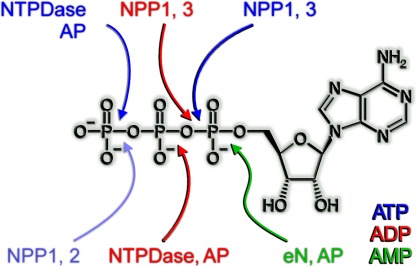
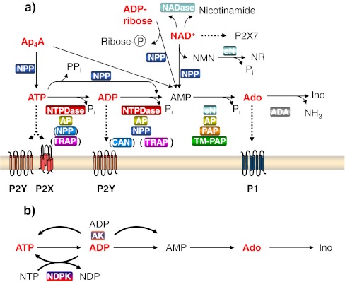
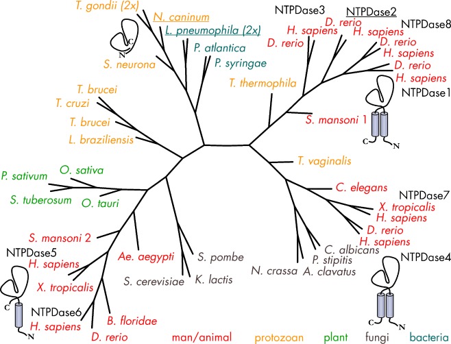
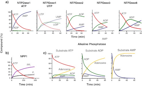
Ecto-nucleotidases play a pivotal role in purinergic signal transmission. They hydrolyze extracellular nucleotides and thus can control their availability at purinergic P2 receptors. They generate extracellular nucleosides for cellular reuptake and salvage via nucleoside transporters of the plasma membrane. The extracellular adenosine formed acts as an agonist of purinergic P1 receptors. They also can produce and hydrolyze extracellular inorganic pyrophosphate that is of major relevance in the control of bone mineralization. This review discusses and compares four major groups of ecto-nucleotidases: the ecto-nucleoside triphosphate diphosphohydrolases, ecto-5'-nucleotidase, ecto-nucleotide pyrophosphatase/phosphodiesterases, and alkaline phosphatases. Only recently and based on crystal structures, detailed information regarding the spatial structures and catalytic mechanisms has become available for members of these four ecto-nucleotidase families. This permits detailed predictions of their catalytic mechanisms and a comparison between the individual enzyme groups. The review focuses on the principal biochemical, cell biological, catalytic, and structural properties of the enzymes and provides brief reference to tissue distribution, and physiological and pathophysiological functions.


Figures

Fig. 1
Fig. 1
Overview of enzyme-specific cleavage sites of individual types of ecto-nucleotidases. Enzymes shown in blue, red, and green accept ATP, ADP and AMP, respectively, as substrates. The cleaved bond is marked by an arrow in the according color. NPPs cleave the same bond in ATP and ADP whereas NTPDases hydrolyze different bonds. The removal of the gamma-phosphate from ATP by NPP1 and NPP2 (pale blue) can be regarded a side reaction of this enzyme class caused by inverted binding of the nucleotide to the active site. See also Fig. 11
Fig. 2
Fig. 2
Principal pathways of extracellular nucleotide metabolism. a Degradation of extracellular nucleotides and purinergic receptor activation with ATP, Ap4A, and NAD+ as examples. Compounds capable of receptor (P2X and P2Y nucleotide receptors or P1 adenosine receptors) activation are indicated in red. Enzymes capable of the specified catalytic reaction are highlighted with a colored background box. ADA ecto-adenosine deaminase, AP alkaline phosphatase, CAN soluble calcium-activated nucleotidase, eN ecto-5′-nucleotidase, NADase NAD-glycohydrolase, NPP ecto-nucleotide pyrophosphatase/phosphodiesterase, NTPDase ecto-nucleoside triphosphate diphosphohydrolase, PAP prostatic acid phosphatase, TM-PAP transmembrane-PAP, TRAP tartrate-resistant acid phosphatase. NAD+ can be hydrolyzed by NPPs to AMP and nicotinamide mononucleotide (NMN) and by NADase to nicotinamide and ADP-ribose. ADP-ribose can in turn be degraded by NPPs to AMP and ribose-5-phosphate. Nicotinamide mononucleotide can be dephosphorylated by eN to nicotinamide riboside (NR). Adenosine (Ado) or inosine (Ino) can be recycled into the cell via specific nucleoside transporters. NAD+ can function as a ligand of some P2 receptors (not indicated), and in murine tissue, it can activate P2X7 receptors as a result of ADP ribosylation. b Ecto-anabolism of nucleotides. At the surface of some cells, ATP can be synthesized extracellularly from ADP via ecto-nucleoside diphosphate kinase (NDPK), whereby another nucleoside triphosphate (NTP) serves as the phosphate donor. In contrast to NDPK, ecto-adenylate kinase (AK) is adenine nucleotide-specific. Depending on mass action, a phosphate can be transferred from one ADP molecule to another, resulting in the formation of ATP and AMP (or vice versa)
Fig. 3
Fig. 3
Radial phylogenetic tree of NTPDases. The tree highlights the clear segregation of vertebrate NTPDases into cell surface-located enzymes which are involved in purinergic signaling (NTPDase1–3 and NTPDase8) and the intracellularly located NTPDase4–7. Although NTPDases are ubiquitous in eukaryotes, cell-surface type forms are probably present in only a few non-vertebrate eukaryotes. The scarcity of bacterial NTPDase genes suggests that they have been acquired by horizontal gene transfer. Amino acid sequences have been aligned with Tcoffee [619]. The tree has been calculated with the program Protdist as included in Bioedit and the visualization was done with the program PhyloDraw. Proteins for which structural data are available have been underscored. GI Accession numbers—A. aegypti: 108878621; Aspergillus clavatus: 121713566; C. elegans: 17539006; Branchiostoma floridae: 210115619; C. albicans: 68480942; Danio rerio NTPDase1: 57525937, NTPDase2: 54261809, NTPDase3: 134133300, NTPDase4: 50539906, NTPDase6: 62955697, NTPDase8: 268837940; Homo sapiens NTPDase1: 1705710, NTPDase2: 45827719, NTPDase3: 4557425, NTPDase4: 3153211, NTPDase5: 3335102, HsNTPDase6: 3335098, NTPDase7: 9623384, NTPDase8: 158705943; Kluyveromyces lactis: 50311623; L. pneumophila: 81377241/gi 81377833; Leishmania braziliensis: 154333055; Neospora caninum: 3298332; Neurospora crassa: 85108997; Pisum sativum: 563612; Oryza sativa: 77548506; Ostreococcus tauri: 308802668; Pichia stipitis: 149389003; Pseudoalteromonas atlantica: 122971633; Pseudomonas syringae pv. tomato: 81730387; S. cerevisiae: 603637; Sarcocystis neurona: 32816824; Schistosoma mansoni 1: 33114187, 2: 114797038; Schizosaccharomyces pombe: 19114359; Solanum tuberosum: 2506931; Tetrahymena thermophila: 118383992; T. gondii: 2499220/gi2499221; Trichomonas vaginalis: 154413345; T. brucei: 72392821; Trypanosoma cruzi: 71414508; Xenopus tropicalis NTPDase5: 301618468, NTPDase7: 62859996
Fig. 4
Fig. 4
Idealized patterns of nucleotide hydrolysis and product formation by select members of the E-NTPDase, E-NPP, and AP families. The formation of ADP from ATP varies between NTPDases (a) and PPi is a major hydrolysis product of NPP1 (b) (the corresponding formation of AMP is not shown). Both types of enzymes produce AMP as final hydrolysis product. On hydrolysis of ATP, AP (c) produces a substrate pattern similar to NTPDase2, NTPDase3, and NTPDase8, except that hydrolysis proceeds to adenosine. Note that only minor amounts of ADP are released on hydrolysis of ATP by NTPDase1. In contrast, hydrolysis of UTP by the same enzyme is progressive with the formation of UDP as intermediate product. Unless indicated otherwise, the initial substrate was ATP. Hydrolysis of AMP by eN (not shown) would follow similar kinetics as those shown for AP. Graphs are modified from the following references: NTPDase1–3 (ATP): heterologous expression of the rat enzymes in CHO cells. Initial substrate concentration 250 or 500 μM [70, 192]; NTPDase1 (UTP), NTPDase8: heterologous expression of mouse enzymes in COS-7 cells. Initial substrate concentration 500 μM [69]. NPP1: Endogenous enzyme expressed by rat C6 glioma cells, presumably NPP1, application of 10 μM [γ-32P]ATP. The pattern of product formation varies, however, with the initial concentration of ATP [369]. The formation of Pi could in part have resulted from the presence of additional ecto-nucleotidases. AP: calf intestinal AP, commercial product from Fermentas Life Sciences, initial substrate concentration 500 μM (Peter Brendel, Frankfurt, unpublished)
Fig. 5
Fig. 5
Structure and mechanism of NTPDases. a Crystal structure of an NTPDase from L. pneumophila (LpNTPDase1) in complex with the substrate analogue AMPPNP. The five ACR regions (numbered) are colored in red. b Model of a membrane-bound NTPDase (RnNTPDase2) based on the complex structure of the rat NTPDase2 ecto-domain with Ca2+ and AMPPNP. Protein regions missing from the crystal structure (due to flexibility in the crystal or being absent in the expression construct [borders indicated by scissors]) are depicted in gray. In addition to the five canonical ACR regions, a sixth region of high sequence conservation among mammalian NTPDases is shown in salmon (numbered 6). This region is symmetry-related to ACR2 and involved in nucleoside binding. A corresponding partial sequence alignment is shown as inset with residues involved in nucleoside binding indicated with triangles. c Superposition of rat NTPDase2 and the NTPDase from L. pneumophila based on domain I (blue). The bacterial NTPDase (gray) represents an open, the mammalian structure (colored) a closed conformation. The relative orientation of the two domains differs by a 14.6° rotation around the axis indicated in purple. In the open state, the substrate analogue adopts a considerably different conformation and does not chelate a metal ion. d Close-up view of the active site of NTPDase2. An extensive network of hydrogen bonds from residues of all six ACRs (numbers in brackets, see text and Table 2 for explanation on the sixth ACR region) functions to orient the substrate and the metal cofactor (yellow sphere) for catalysis. Hydrolysis proceeds via activation of a water molecule (orange) by the catalytic base E165
Fig. 6
Fig. 6
Schematic representation of the postulated reaction mechanism of NTPDase-mediated NTP (R = NMP) or NDP (R = nucleoside) hydrolysis. a Activation of the nucleophilic water by E165 and subsequent inline nucleophilic attack on the terminal phosphate. The negative charge of the phosphate groups is reduced by complexation of the divalent metal cation. A positively charged H50 may additionally draw partial negative charge from the terminal to the penultimate phosphate group, lowering the activation barrier. b Collapse of the trigonal bipyramidal transition state. The negative charge of the transition state is stabilized by proton-donating hydrogen bonds from the phosphate-binding loops (e.g., S48, A205). Additional hydrogen bonds may exist. c Product release. H50 may be responsible for protonation of the leaving group. d Reconstitution of the active site
Fig. 7
Fig. 7
Radial phylogenetic tree and substrate specificity of members of the 5′-nucleotidase family. Availability of structural data is indicated by underscores. Functional data are available for some purified nucleotidases. Accepted substrates are shown in black. No turnover could be detected with substances shown in red. Mammalian eN occurs in dimeric form, is membrane-attached via a GPI anchor. Bacterial 5′-nucleotidases are monomeric. Membrane attachment via a lipid anchor has been reported for Vibrio parahaemolyticus [620] and the 5′-nucleotidase from R. microplus seems to be membrane-bound via a GPI anchor [621]. No functional data are available for 5′-nucleotidases from yeasts and fungi which are phylogenetically distantly related to those of bacteria and animals. Amino acid sequences have been aligned with ClustalW and the tree was calculated with ProML. GI accession numbers and references for functional studies—A. aegypti (mosquito): gi556272 [293]; Bos taurus: gi285642; C. albicans: 68477828; D. rerio: 32766695; Discopyge ommata (electric ray): 62772; E. coli (ushA): 2506086; E. coli CFT073 (c4898): 26250712; Glossina morsitans morsitans (Tse tse fly): 14488055 [622]; Homo sapiens: 4505467; Kluyveromyces lactis: 50303513; L. longipalpis (sandfly): 4887100; Mus musculus: 6754900; Ornithodoros savignyi (soft tick): 152207619 [623]; R. microplus (cattle tick): 1737443; S. aureus: 88193920; T. thermophilus: 55981297; Treponema pallidum: 5902689; T. infestans: 34481604 [295]; Trichinella spiralis: 22656349 [624]; Vibrio parahaemolyticus: 217191. UDPG, UDP-glucose
Fig. 8
Fig. 8
Crystal structure of Ec5NT. a Open form of the enzyme. The N-terminal domain (yellow) binds the two metal ions (light blue). An ATP molecule (blue) is complexed to the substrate-binding pocket of the C-terminal domain (red). b Closed form of the enzyme in complex with AMPCP. The non-hydrolysable ADP analogue binds between the two domains. The nucleoside moiety of the substrate analogue is bound to the same binding pocket as in a, which is rotated together with the C-terminal domain by ∼96° around the axis shown in green. The bending residues, i.e., the region of the protein which accommodates the rotation of the rigid domains, are depicted in green. c Binding mode of the substrate analogue inhibitor α,β-methylene-ADP to the closed form of Ec5NT (stereo view). The residues which are not conserved in eN are depicted in broken lines. The residue labels refer to the human enzyme. The loop shown in green is predicted to be shorter in the ecto-enzymes. The coloring of the protein domains is the same as in a. d Scheme of the interactions involved in inhibitor binding. The upper label for each residue refers to human eN and the bottom label to Ec5NT
Fig. 9
Fig. 9
Scheme of the dimetal center and proposed structure of the Michaelis complex for eN catalysis. The binding mode of the substrate’s terminal phosphate group is supported by the binding mode of the inhibitor α,β-methylene-ADP to Ec5NT. Residue labels refer to human eN. All residues except for N245 are conserved in the E. coli enzyme. Therefore, the coordination of N245 via a water molecule to metal ion 1 is hypothetical. In the bacterial enzyme, N245 is replaced by a glutamine, which is directly coordinated to the metal ion
Fig. 10
Fig. 10
Radial phylogenetic tree and membrane topology of NPP-type ecto-enzymes. The tree highlights the separation between the multidomain NPP1–3 and NPP4-7, which consist of only the catalytic domain. Separation into the seven different NPPs has occurred early in evolution. Included is the bacterial NPP from X. axonopodis pv. citri, for which structural data are available. NPP-type enzymes are ubiquitous in nature. Amino acid sequences have been aligned with ClustalW and the tree was calculated with ProML. GI Accession numbers (clockwise): D. rerio NPP2: 82187289, NPP4: 62955749, NPP5: 113462025, NPP6: 61806626; H. sapiens NPP1: 23503088, NPP2: 290457674, NPP3: 206729860, NPP4: 172045555, NPP5: 50401201, NPP6: 108935979, NPP7: 134047772; X. axonopodis pv. citri: 21243551; X. laevis NPP3: 254946550; X. tropicalis NPP1: 254946558, NPP7: 296010817
Fig. 11
Fig. 11
Substrate specificity of NPP-type ecto-enzymes. Catalysis is thought to occur by nucleophilic attack of the Thr alkoxide onto the boxed phosphorus atom, so that a covalent intermediate with the R′ group is formed. In a second step, the covalent intermediate is hydrolyzed by nucleophilic attack from a Zn2+-activated water. To some of the enzymes, the substrates may bind in both orientations and this is also reflected by their ability to hydrolyze dinucleotides (e.g., NPP1 and NPP3). NPP2 and NPP6/7 are active towards the same class of substrates. Due to swapped specificities in the R and R′ binding sites, the substrates bind in an inverted position resulting in the generation of different hydrolysis products. The scheme is based on data presented in the following papers—NPP1: [351, 353, 412, 453, 625]; NPP2: [351, 353, 377, 380, 403, 406]; NPP3: [351]; NPP6: [388]; NPP7: [389, 626]. GPC glycerophosphorylcholine, LPC lysophosphatidylcholine, pNPPC p-nitrophenyl phosphorylcholine, SPC sphingosylphosphorylcholine
Fig. 12
Fig. 12
Structure of NPP-type enzymes. a Domain organization of NPPs: NPP1–3 are composed of two SMB domains, the catalytic domain and a nuclease-like domain. In contrast, NPP4–7 lack any additional non-catalytic domains. An NPP-specific insertion into the AP fold “CAP” is shown in a lighter color. The position of the NPP2-specific deletion required for formation of the two LPA-binding sites is indicated. b Ribbon/surface representation of mouse NPP2 and NPP from X. axonopodis pv. citri (Xac) with an identical view on the active site. The bound product molecules LPA and AMP are shown as sticks and metal ions (2 × Zn2+, Ca2+, Na+, K+) as spheres. The WPG loop of Xac NPP that is missing in NPP2 (see d) is shown in yellow. c Surface representation of NPP2 along the LPA channel showing the intricate arrangement of the four domains, the linker, and the long lasso regions. N-linked glycosylation sites are shown as sticks and the structurally important N524 glycan is labeled. The eye indicates the approximate view in b. d Surface representation of NPP2 with a longitudinal section along the hydrophobic LPA-binding channel. Only one of the Zn2+ ions is visible. One LPA molecule (LPA1) is bound to the active site and the hydrophobic pocket. The second LPA molecule (partial model, LPA2) binds in the hydrophobic channel more distal to the active site. In all other NPP-type enzymes, these two binding sites are occluded by an amino acid stretch consisting of a highly conserved WPG motif at the beginning and a tyrosine at the end that is involved in binding the base of nucleotide substrates. The corresponding loop region and active site-bound AMP of Xac NPP is shown in green and magenta sticks, respectively. LPA lysophosphatidic acid, SMB somatomedin B
Fig. 13
Fig. 13
Comparison of the active site structures of human placental AP (PLAP) and human NPP1. The active site structure of human NPP1 differs from PLAP mainly by the absence of the magnesium ion binding site, the replacement of Arg-166 by Asn-277, and the presence of a substrate specificity site formed by Tyr-371, Phe-321, Phe-257, and Leu-290
Fig. 14
Fig. 14
Radial phylogenetic tree and substrate specificity of APs. Genes for canonical APs are ubiquitous but have not been cloned from plants. The tree highlights the closer relation of tissue nonspecific APs (TNAP) to those found in bacteria, archaea, and invertebrates. The divergence of the other human APs has occurred rather late in evolution. Availability of structural data is indicated by underscores. Amino acid sequences have been aligned with ClustalW and the tree was calculated with ProML. GI accession numbers—Antarctic bacterium TAB5: 7327837; D. rerio AP: 41055949; intestinal AP: 62122905, intestinal AP2: 68448521, E. coli: 581187; H. salinarum: 167728700; H. sapiens germ cell AP (GCAP): 110347479, intestinal AP (IAP): 157266292, placental AP (PLAP): 94721246, TNAP: 116734717; Mus musculus embryonic AP (EAP): 7327837, TNAP: 160333226, intestinal AP (IAP): 110347479; Pandalus borealis (shrimp): 13539555; Shewanella sp.: 19071967; Vibrio sp.: 243065523
Fig. 15
Fig. 15
Crystal structure of human placental AP (PLAP). a Stereo view of the fold of PLAP. The view is perpendicular to the twofold molecular axis relating the two monomers of the dimeric protein. Monomer A is colored in red and the other monomer B in beige. These regions correspond to the core part of the monomer that is conserved with the bacterial APs. Additional domains in the mammalian protein are colored as follows. The crown domain is shown in light blue (monomer A) and blue (monomer B). The metal-binding domain is indicated in yellow. The N-terminal helices (green, monomer A and orange, monomer B) form contacts to the neighboring monomer. The zinc ions of the active site are shown in green and the magnesium ions in white. The glycosylation sites are indicated in cyan. The C-terminal residue (479) of the X-ray structure is shown in red (large spheres) to indicate a position close to Asp484, to which the GPI anchor is attached for immobilization of the protein to the cell membrane. A phenylalanine ligand bound to a peripheral binding site is shown in red sticks (hidden behind the protein in monomer A). Generated from pdb ids 1EW2 and 1ZEF. b Stereo view of the active site of PLAP. The zinc ions are shown in green and the magnesium ion is depicted as a gray sphere. The coordination bonds are shown as broken lines. Tyr367, depicted with yellow carbon atoms, belongs to the neighboring subunit of the dimeric protein. The interactions between Arg166 and the bound phosphate ion are indicated as broken lines. Generated from PDB id 1EW2. c Inhibitory binding mode of l-Phe in the active site of PLAP. The zinc ions are shown in green and the magnesium ion is depicted as a gray sphere. Coordination bonds and hydrogen bonding interactions are shown as broken lines. The bound phenylalanine inhibitor is depicted with yellow carbon atoms. Generated from PDB id 3MK2. See the electronic form of the article for a colored version of this figure
Fig. 16
Fig. 16
Proposed reaction mechanism of AP [586]. a Scheme of the active site in the unliganded state. bd Michaelis complex, transition state, and product complex, respectively, of the formation of the phosphorylated serine intermediate. eg Dephosphorylation of the intermediate. The residue numbers refer to human placental AP (PLAP)

References

    1. Burnstock G. Physiology and pathophysiology of purinergic neurotransmission. Physiol Rev. 2007;87:659–797. doi: 10.1152/physrev.00043.2006. - DOI - PubMed
    1. Zimmermann H, Mishra SK, Shukla V, Langer D, Gampe K, Grimm I, Delic J, Braun N. Ecto-nucleotidases, molecular properties and functional impact. An R Acad Nac Farm. 2007;73:537–566.
    1. Zimmermann H. ATP and acetylcholine, equal brethren. Neurochem Int. 2008;52:634–648. doi: 10.1016/j.neuint.2007.09.004. - DOI - PubMed
    1. Burnstock G. Pathophysiology and therapeutic potential of purinergic signaling. Pharmacol Rev. 2006;58:58–86. doi: 10.1124/pr.58.1.5. - DOI - PubMed
    1. King AE, Ackley MA, Cass CE, Young JD, Baldwin SA. Nucleoside transporters: from scavengers to novel therapeutic targets. Trends Pharm Sci. 2006;27:416–425. doi: 10.1016/j.tips.2006.06.004. - DOI - PubMed

Publication types

MeSH terms

LinkOut - more resources