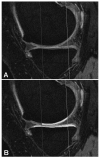
Cartilage thickening in early radiographic knee osteoarthritis: a within-person, between-knee comparison
- PMID: 22556039
- PMCID: PMC3429643
- DOI: 10.1002/acr.21719
Cartilage thickening in early radiographic knee osteoarthritis: a within-person, between-knee comparison
Abstract
Objective: To determine whether the presence of definite osteophytes (in the absence of joint space narrowing [JSN]) on radiographs is associated with (subregional) increases in cartilage thickness in a within-person, between-knee cross-sectional comparison of participants in the Osteoarthritis Initiative. Based on previous results, the external weight-bearing medial femoral condyle (ecMF) and external weight-bearing lateral femoral condyle (ecLF) subregions were selected as primary end points.
Methods: Both knees of 61 Osteoarthritis Initiative participants (n = 4,796) displayed definite tibial or femoral marginal osteophytes and no JSN in 1 knee, and no signs of radiographic osteoarthritis (OA) in the contralateral knee; this was confirmed by an expert central reader. In these participants, cartilage thickness was measured in 16 femorotibial subregions of each knee, based on sagittal double-echo steady-state with water excitation magnetic resonance images. Location-specific joint space width from fixed-flexion radiographs was determined using dedicated software. Location-specific associations of osteophytes with cartilage thickness were evaluated using paired t-tests and mixed-effects models.
Results: Of the 61 participants, 48% had only medial osteophytes, 36% only lateral osteophytes, and 16% bicompartmental osteophytes. The knees with osteophytes had significantly thicker cartilage than contralateral knees without osteophytes in the ecMF (mean ± SD +71 ± 223 μmoles, equivalent to an increase of +5.5%; P = 0.015) and ecLF (mean ± SD +64 ± 195 μmoles, +4.1%; P = 0.013). No significant differences between knees were noted in other subregions or in joint space width. Cartilage thickness in the ecMF and ecLF was significantly associated with tibial osteophytes in the same (medial or lateral) compartment (P = 0.003).
Conclusion: The knees with early radiographic OA display thicker cartilage than (contralateral) knees without radiographic findings of OA, specifically in the external femoral subregions of compartments with marginal osteophytes.
Copyright © 2012 by the American College of Rheumatology.
Figures


Similar articles
-
Longitudinal (one-year) change in cartilage thickness in knees with early knee osteoarthritis: A within-person between-knee comparison.Arthritis Care Res (Hoboken). 2014 Apr;66(4):636-41. doi: 10.1002/acr.22172. Arthritis Care Res (Hoboken). 2014. PMID: 24106150
-
Hand joint space narrowing and osteophytes are associated with magnetic resonance imaging-defined knee cartilage thickness and radiographic knee osteoarthritis: data from the Osteoarthritis Initiative.J Rheumatol. 2012 Jan;39(1):161-6. doi: 10.3899/jrheum.110603. Epub 2011 Nov 1. J Rheumatol. 2012. PMID: 22045837
-
Magnetic resonance imaging-based cartilage loss in painful contralateral knees with and without radiographic joint space narrowing: Data from the Osteoarthritis Initiative.Arthritis Rheum. 2009 Sep 15;61(9):1218-25. doi: 10.1002/art.24791. Arthritis Rheum. 2009. PMID: 19714595 Free PMC article.
-
Imaging of cartilage and bone: promises and pitfalls in clinical trials of osteoarthritis.Osteoarthritis Cartilage. 2014 Oct;22(10):1516-32. doi: 10.1016/j.joca.2014.06.023. Osteoarthritis Cartilage. 2014. PMID: 25278061 Free PMC article. Review.
-
An OMERACT reliability exercise of inflammatory and structural abnormalities in patients with knee osteoarthritis using ultrasound assessment.Ann Rheum Dis. 2016 May;75(5):842-6. doi: 10.1136/annrheumdis-2014-206774. Epub 2015 Apr 22. Ann Rheum Dis. 2016. PMID: 25902788
Cited by
-
Morphological and histological features of thicker cartilage at the posterior medial femoral condyle in advanced knee osteoarthritis.Osteoarthr Cartil Open. 2024 Jul 11;6(3):100502. doi: 10.1016/j.ocarto.2024.100502. eCollection 2024 Sep. Osteoarthr Cartil Open. 2024. PMID: 39114819 Free PMC article.
-
Baseline-to-loaded changes in regional tibial cartilage thickness, T1ρ and T2: Utilization of an MRI compatible loading device.J Orthop Res. 2024 Dec;42(12):2646-2658. doi: 10.1002/jor.25956. Epub 2024 Aug 23. J Orthop Res. 2024. PMID: 39177306
-
Effects of Joint Immobilization and Treadmill Exercise on Articular Cartilage After ACL Reconstruction in Rats.Orthop J Sports Med. 2022 Oct 17;10(10):23259671221123543. doi: 10.1177/23259671221123543. eCollection 2022 Oct. Orthop J Sports Med. 2022. PMID: 36276424 Free PMC article.
-
Ultrasonographic Assessment of Femoral Cartilage in Individuals With Anterior Cruciate Ligament Reconstruction: A Case-Control Study.J Athl Train. 2018 Nov;53(11):1082-1088. doi: 10.4085/1062-6050-376-17. Epub 2019 Jan 7. J Athl Train. 2018. PMID: 30615493 Free PMC article.
-
Association of age, sex and BMI with the rate of change in tibial cartilage volume: a 10.7-year longitudinal cohort study.Arthritis Res Ther. 2019 Dec 9;21(1):273. doi: 10.1186/s13075-019-2063-z. Arthritis Res Ther. 2019. PMID: 31818318 Free PMC article.
References
-
- Watson PJ, Carpenter TA, Hall LD, et al. Cartilage swelling and loss in a spontaneous model of osteoarthritis visualized by magnetic resonance imaging. Osteoarthritis Cartilage. 1996;4:197–207. - PubMed
-
- Calvo E, Palacios I, Delgado E, et al. Histopathological correlation of cartilage swelling detected by magnetic resonance imaging in early experimental osteoarthritis. Osteoarthritis Cartilage. 2004;12:878–86. - PubMed
-
- Calvo E, Palacios I, Delgado E, et al. High-resolution MRI detects cartilage swelling at the early stages of experimental osteoarthritis. Osteoarthritis Cartilage. 2001;9:463–72. - PubMed
-
- Tessier JJ, Bowyer J, Brownrigg NJ, et al. Characterisation of the guinea pig model of osteoarthritis by in vivo three-dimensional magnetic resonance imaging. Osteoarthritis Cartilage. 2003;11:845–53. - PubMed
Publication types
MeSH terms
Grants and funding
LinkOut - more resources
Full Text Sources
Other Literature Sources

