. 2012 Jul 1;72(13):3187-95.
doi: 10.1158/0008-5472.CAN-12-0534. Epub 2012 May 3.

Loss of fibroblast HIF-1α accelerates tumorigenesis

Affiliations

Loss of fibroblast HIF-1α accelerates tumorigenesis

Jung-whan Kim et al. Cancer Res. .

Abstract

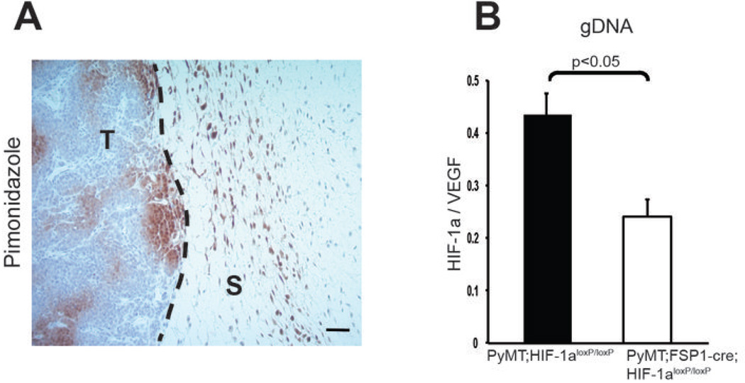
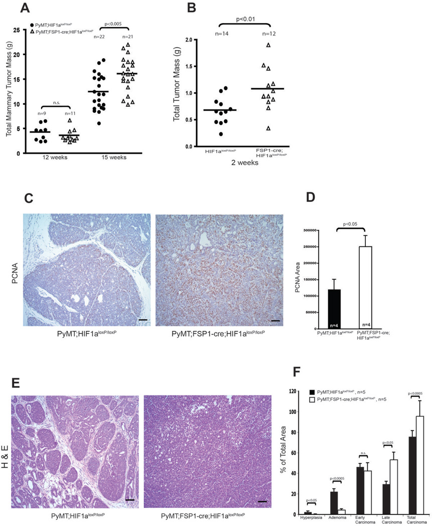
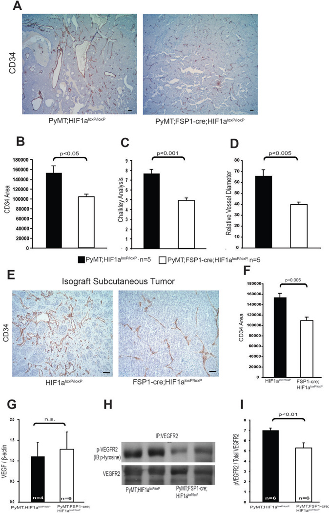
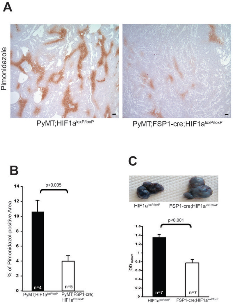
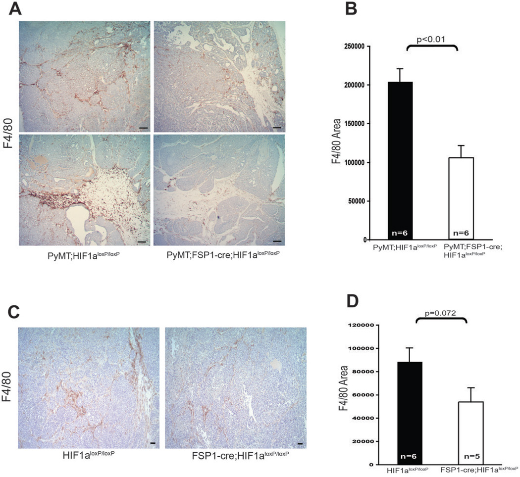
Solid tumors consist of malignant cells and associated stromal components, including fibroblastic cells that contribute to tumor growth and progression. Although tumor fibrosis and aberrant vascularization contribute to the hypoxia often found in advanced tumors, the contribution of hypoxic signaling within tumor-associated fibroblasts to tumorigenesis remains unknown. In this study, we used a fibroblast-specific promoter to create mice in which key hypoxia regulatory genes, including VHL, HIF-1α, HIF-2α, and VEGF-A, were knocked out specifically in tumor stromal fibroblasts. We found that loss of HIF-1α and its target gene VEGF-A accelerated tumor growth in murine model of mammary cancer. HIF-1α and VEGF-A loss also led to a reduction in vascular density and myeloid cell infiltration, which correlated with improved tumor perfusion. Together, our findings indicate that the fibroblast HIF-1α response is a critical component of tumor vascularization.


Conflict of interest statement

The authors declare no conflict of interest

Figures

Figure 1
Figure 1. Targeted Deletion of HIF-1α in stromal fibroblasts
(A) Hypoxic areas (brown) in transgenically induced mammary tumors were detected by immunostaining for the hypoxia marker of bioreductivity, pimonidazole; scale bar, 100 µm. T, tumor; S, stroma. (B) Genomic DNA extracted from stromal fibroblasts isolated from WT and fibroblast HIF-1α null mammary tumors by culturing on petri dish were analyzed for the expression of HIF-1α. Genomic DNA level was normalized to VEGF-A genomic sequence as a non-deleted control for gene dosage.
Figure 2
Figure 2. Deletion of Stromal Fibroblast HIF-1α Promotes Mammary Tumor Growth and Progression
(A) Total mammary tumor mass of WT (PyMT;Hif-1αloxp/loxp) and fibroblast HIF-1α null (PyMT;Fsp-cre;Hif-1αloxp/loxp) mice measured at the indicated age. (B) Total isograft tumor burden at 2 weeks post-subcutaneous injection of tumor cells into WT (Hif-1αloxp/loxp) and fibroblast HIF-1α null (Fsp-cre;Hif-1αloxp/loxp) mice. (C) Immunostaining for PCNA in tumor sections from 15-week-old WT (PyMT;Hif-1αloxp/loxp) and fibroblast HIF-1α null (PyMT;Fsp-cre;Hif-1αloxp/loxp) mice; scale bars: 100µm. (D) Quantification of PCNA-positive areas in sections represented in Fig. 2C. (E) Representative images of H&E stained mammary tumors collected from 15-week-old WT (PyMT;Hif-1αloxp/loxp) and fibroblast HIF-1α null (PyMT;Fsp-cre;Hif-1αloxp/loxp) mice; scale bars: 100µm. (F) Histological stage distribution of mammary tumors from 15-week-old WT (PyMT;Hif-1αloxp/loxp) and fibroblast HIF-1α null (PyMT;Fsp-cre; Hif-1αloxp/loxp) mice.
Figure 3
Figure 3. Ablation of HIF-1α in fibroblasts reduces tumor vascularization
(A) Tumor vasculature was visualized by CD34 immunostaining of tumor sections collected from 15-week-old WT (PyMT;Hif-1αloxp/loxp) and fibroblast HIF-1α null (PyMT;Fsp-cre;Hif-1αloxp/loxp) mice; scale bars: 100µm. (B) Quantification of CD34-positive stained areas from the tumors in Fig 3A. (C) Quantification of blood vessel density determined by Chalkley analysis of the tumors described for Fig. 3A. (D) Quantification of blood vessel diameter in the tumors described for Fig. 3A. (E) Representative sections of CD34-stained isograft subcutaneous tumors collected 2 weeks post-injection of syngeneic mammary tumor cells in WT (PyMT;Hif-1αloxp/loxp) and fibroblast HIF-1α null (PyMT;Fsp-cre;Hif-1αloxp/loxp) mice; scale bars: 100µm. (F) Quantification of CD34-positive stained area in tumors described in Fig. 3E. (G) Quantification of VEGF-A mRNA expression in mammary tumors. Values represent mean ratio of VEGF-A mRNA to β-actin mRNA ± SEM. (H) Representative immunoblot analysis of phosphotyrosine presence in immunoprecipitated VEGFR2, and total VEGFR2 in mammary tumor lysates. (I) Quantification of phosphotyrosine and VEGFR2 by measuring photon emission in the blots described in Fig. 3H. Values represent the mean ratio of phosphotyrosine to total VEGFR2 photon emission intensities ± SEM.
Figure 4
Figure 4. Targeted Deletion of HIF-1α in Fibroblasts reduces hypoxia
(A) Hypoxic areas in mammary tumor sections from 15-week-old WT (PyMT;Hif-1αloxp/loxp) and fibroblast HIF-1α null (PyMT;Fsp-cre;Hif-1αloxp/loxp) mice were visualized by immunostaining for the hypoxia marker pimonidazole; scale bar, 100 µm. (B) Quantification of pimonidazole staining in mammary tumors referred to in Fig. 4A. (C) Upper: Evans blue dye extravasation in subcutaneous tumors collected from WT (PyMT;Hif-1αloxp/loxp) and fibroblast HIF-1α null (PyMT;Fsp-cre; Hif-1αloxp/loxp) mice at 2 weeks post-injection of tumor cells. Lower: Quantification of extravasated Evans blue dye.
Figure 5
Figure 5. Fibroblast Hif-1α Deletion Attenuates Macrophage Infiltration in Mammary Tumors
(A) Representative mammary tumor sections from 15-week-old WT (PyMT;Hif-1αloxp/loxp) and fibroblast HIF-1α null (PyMT;Fsp-cre;Hif-1αloxp/loxp) mice, stained for the macrophage marker F4/80; scale bars: 100µm. (B) Quantification of F4/80-stained area in mammary tumors as in Fig. 5A. (C) Representative sections from subcutaneous tumors at 2 weeks post-injection of tumor cells in WT (PyMT;Hif-1αloxp/loxp) and fibroblast HIF-1α null (PyMT;Fsp-cre; Hif-1αloxp/loxp) mice stained for F4/80; scale bars: 100µm. (D) Quantification of F4/80 stained area in tumors described in Fig. 5C.
Figure 6
Figure 6. Deletion of VEGF-A in Fibroblasts Promotes Tumor Growth in syngeneic isografts
(A) Total isograft tumor mass at 2 weeks post-subcutaneous injection of tumor cells into WT (Vegfaloxp/loxp) and fibroblast VEGF-Fibroblast A null (Fsp-cre;Vegfaloxp/loxp) mice. (B) Representative image of isograft subcutaneous tumors harvested from WT (Vegfaloxp/loxp) and fibroblast VEGF-A null (Fsp-cre;Vegfaloxp/loxp) mice at 2 weeks post-injection of tumor cells. (C) Representative tumor sections from 15-week-old WT (Vegfaloxp/loxp) and fibroblast VEGF-A null (Fsp-cre;Vegfaloxp/loxp) mice stained for CD34 (upper panels) and F4/80 (bottom panels); scale bars: 100µm. (D) Quantification of CD34- (left) and F4/80- (right) stained area in tumors described in Fig. 6C.

References

    1. Egeblad M, Nakasone ES, Werb Z. Tumors as organs: complex tissues that interface with the entire organism. Developmental cell. 2010;18:884–901. - PMC - PubMed
    1. Bissell MJ, Radisky D. Putting tumours in context. Nature reviews Cancer. 2001;1:46–54. - PMC - PubMed
    1. Polyak K, Haviv I, Campbell IG. Co-evolution of tumor cells and their microenvironment. Trends in genetics : TIG. 2009;25:30–38. - PubMed
    1. Kalluri R, Zeisberg M. Fibroblasts in cancer. Nature reviews Cancer. 2006;6:392–401. - PubMed
    1. Bhowmick NA, Neilson EG, Moses HL. Stromal fibroblasts in cancer initiation and progression. Nature. 2004;432:332–337. - PMC - PubMed

Publication types

MeSH terms

Substances